Did you know that ACL and PCL tears create completely different patterns of knee instability despite occurring in the same joint? The anterior cruciate ligament (ACL) and posterior cruciate ligament (PCL) form an X-shape inside your knee joint, stabilising the connection between your thighbone and shinbone. ACL tears occur when the ligament stretches beyond its capacity during sudden pivoting or landing movements, while PCL tears typically result from direct impact to the front of the knee when it’s bent.
Anatomy and Function of Knee Ligaments
The ACL runs diagonally from the back of the femur to the front of the tibia, preventing the shinbone from sliding forward. The PCL connects the front of the femur to the back of the tibia, preventing backward displacement of the tibia.
The ACL activates primarily during acceleration, deceleration, and rotational movements. When a person plants their foot and pivots during sports, the ACL experiences maximum tension. The PCL handles different stresses—it engages most when the knee bends beyond 90 degrees or when force pushes the shinbone backwards relative to the thighbone.
These ligaments work together with the medial and lateral collateral ligaments to maintain knee stability through the full range of motion. During walking, both ligaments share the load, but their individual contributions vary with knee position and the forces applied.
Mechanism of Injury
ACL tears commonly occur through non-contact mechanisms during sports that involve cutting, jumping, and sudden direction changes. The typical scenario involves planting the foot while the body rotates, creating a twisting force that overwhelms the ligament. Basketball players experience this when landing from a jump with their knee extended and rotated. Soccer players tear their ACL when rapidly changing direction with a planted foot.
PCL injuries require different forces—usually a direct blow to the front of the knee. Dashboard injuries in car accidents represent a common PCL tear mechanism, where the bent knee strikes the dashboard, driving the tibia backwards. In sports, PCL tears happen when athletes fall onto a flexed knee with the foot pointing downward, concentrating force through the shinbone.
Contact sports create scenarios for both injuries. A tackle hitting the outside of a planted leg can tear the ACL through forced inward bending and rotation. The same tackle to a bent knee might rupture the PCL if it drives the lower leg backwards. The injury mechanism often determines which ligament tears and whether other structures sustain damage.
Clinical Examination and Diagnosis
Orthopaedic surgeons use physical tests to differentiate ACL from PCL tears. The Lachman test evaluates ACL integrity by pulling the tibia forward with the knee bent at 30 degrees. A positive test shows excessive forward movement compared to the uninjured side. The anterior drawer test assesses the knee at 90 degrees of flexion.
PCL examination relies on the posterior drawer test, in which the surgeon pushes the tibia posteriorly with the knee bent. The posterior sag sign provides another diagnostic clue: when both knees are bent to 90 degrees while lying down, the PCL-injured side shows the tibia sagging backwards relative to the femur.
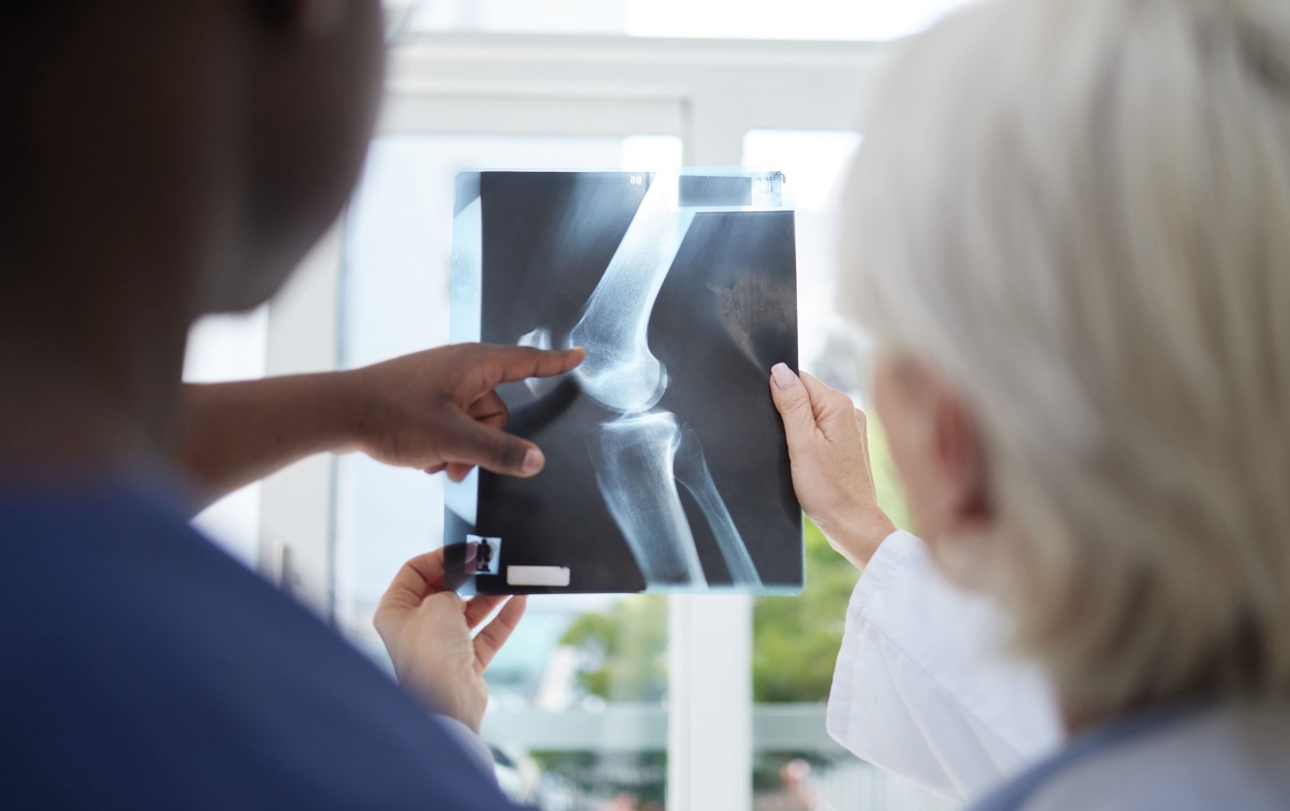
MRI scanning confirms the clinical diagnosis and reveals associated injuries. ACL tears appear as discontinuous or wavy ligament fibres on sagittal images. PCL tears show similar disruption, but in the posterior knee compartment. MRI also identifies bone bruising patterns—ACL injuries typically bruise the lateral femoral condyle and posterior lateral tibia, while PCL injuries affect the anterior tibia.
Treatment Approaches
ACL reconstruction is a treatment option for active individuals who want to return to pivoting sports. For patients considering acl surgery in Singapore, an orthopaedic surgeon will usually assess the severity of the tear, knee stability, activity level, and any associated meniscus or cartilage injuries before recommending surgery. Surgeons harvest tendon tissue from your patellar tendon, hamstrings, or quadriceps to create a new ligament. The procedure involves drilling tunnels through the femur and tibia, threading the graft through these tunnels, and securing it with screws or buttons. Surgery timing matters—operating immediately after injury increases the risk of stiffness, so surgeons typically wait for swelling to subside.
PCL treatment follows different principles because this ligament may have an improved healing potential than the ACL. Many partial PCL tears and some complete tears heal adequately with conservative treatment. The rehabilitation protocol emphasises quadriceps strengthening to compensate for PCL deficiency. When surgery becomes necessary—typically for combined ligament injuries or failed conservative treatment—the reconstruction technique mirrors ACL surgery but uses different tunnel positions.
Post-surgical rehabilitation progresses differently between ligaments. ACL rehabilitation limits early knee flexion to protect the graft, while PCL protocols restrict posterior tibial translation. Return to sports typically takes several months after ACL reconstruction and less time after isolated PCL reconstruction, though combined injuries require more extended recovery periods.
Recovery Timeline and Rehabilitation
ACL rehabilitation follows distinct phases over 9-12 months. The first two weeks focus on reducing swelling and regaining knee extension. Weeks 2-6 introduce progressive weight-bearing and range-of-motion exercises. By month 3, patients begin straight-line running if they’ve achieved full extension and adequate quadriceps strength. Sport-specific training starts around month 6, with return to competition after passing functional tests at 9-12 months.
PCL rehabilitation emphasises quadriceps activation from day one. The quadriceps muscles pull the tibia forward, compensating for PCL deficiency. Early exercises avoid hamstring activation, which draws the tibia backwards. Weight bearing begins immediately with the knee in slight flexion. Running typically resumes by month 3, earlier than ACL protocols, because forward-backwards stability affects straight-line activities less than rotational stability.
Both rehabilitation programs require achieving specific milestones before progression. Full knee extension, minimal swelling, and adequate muscle activation allow advancement to the next phase. Functional tests, including single-leg hop tests, agility drills, and strength measurements, guide return-to-sport decisions.
Long-term Implications
ACL-deficient knees develop characteristic degenerative patterns over time. The excessive forward tibial translation damages the meniscus, particularly the posterior horn of the medial meniscus. Cartilage wear occurs in predictable locations—the medial compartment and patellofemoral joint show accelerated degeneration. Even after reconstruction, there is an increased arthritis risk of arthritis compared to uninjured knees. In severe cases of long-term knee degeneration, treatment may shift from ligament reconstruction to joint-focused procedures such as total knee replacement in Singapore, depending on the extent of cartilage damage and symptoms.
PCL deficiency creates different wear patterns. The posterior tibial subluxation increases contact pressures in the medial compartment and patellofemoral joint. Unlike ACL injuries, PCL deficiency particularly affects the cartilage behind the kneecap. Many patients develop anterior knee pain years after the initial injury.
Activity modification may help both conditions. ACL-deficient patients often continue straight-line activities but may experience challenges with cutting sports. PCL-deficient individuals adapt to most activities but may report difficulty with prolonged kneeling or squatting.
What an Orthopaedic Surgeon Says
The decision between surgery and conservative treatment depends on multiple factors beyond just which ligament is torn. A knee specialist in Singapore can assess the injury pattern, activity demands, and recovery goals to determine whether rehabilitation, bracing, or reconstruction is more appropriate. A competitive athlete with an ACL tear almost always needs reconstruction to return safely to their sport. However, many PCL tears can be treated successfully without surgery, especially in patients whose activities don’t stress the posterior structures.
The timing of intervention matters significantly. Rushing into ACL surgery before regaining motion leads to stiffness complications. Conversely, delaying too long allows muscle atrophy and potentially additional meniscus damage. Each patient’s injury pattern, activity level, and personal goals shape the treatment recommendation.
Current reconstruction techniques have substantially improved outcomes. Surgeons now position tunnels more anatomically and can address associated injuries simultaneously. Still, no reconstruction perfectly replicates the native ligament. That’s why injury prevention through neuromuscular training is emphasised, particularly for young athletes in high-risk sports.
Commonly Asked Questions
How can I tell if I’ve torn my ACL or PCL without an MRI?
The injury mechanism provides clues—non-contact twisting injuries suggest ACL tears, while dashboard injuries or falls onto bent knees indicate PCL damage. ACL tears cause immediate pain and rapid swelling, while PCL injuries develop symptoms gradually. Your knee instability pattern also differs: ACL tears cause giving way during pivoting, while PCL tears create problems with deceleration and stairs.
Can these ligaments heal without surgery?
The ACL has limited healing capacity due to poor blood supply and the synovial environment. Complete ACL tears rarely heal to provide stability. PCL tears, especially partial tears, can heal with rehabilitation. The PCL’s improved blood supply and extrasynovial position create a more favourable healing environment. Many patients with isolated PCL tears achieve functional improvement with conservative treatment.
What activities can I do with a torn ACL or PCL while waiting for surgery?
With ACL tears, straight-line activities like cycling, swimming, and elliptical training remain safe options. Avoid any cutting, pivoting, or jumping activities. PCL tears allow more activity variety, but limit deep squatting, kneeling, and high-impact deceleration. Both injuries benefit from maintaining quadriceps and hip strength during the waiting period.
Will I develop arthritis after ligament reconstruction?
Reconstructed knees show higher arthritis rates than uninjured knees in long-term studies. Meniscus damage at the time of injury represents a predictor of future arthritis. Delaying surgery allows additional meniscus damage, while early treatment preserves joint structures. Modern anatomic reconstruction techniques may reduce the risk of arthritis compared to older methods.
How do I know when I’m ready to return to sports?
Return-to-sport criteria include both time-based and functional markers. Your operated leg may achieve a similar level of strength to the uninjured side. Single-leg hop tests must reach symmetry across four different tests. Psychological readiness assessments help identify fear of reinjury. You can complete sport-specific training without pain, swelling, or episodes of instability.

Experiencing Knee Pain or Injury?
Get a Personalised Treatment Plan
Find relief with our hip & knee specialists.
Make An EnquiryConclusion
ACL reconstruction typically requires 9-12 months for sports return, while PCL injuries often heal with conservative treatment and shorter rehabilitation periods. Both injuries need a prompt, accurate diagnosis to prevent long-term cartilage damage. Modern surgical techniques and structured rehabilitation protocols optimise outcomes when conservative treatment proves insufficient.
If you’re experiencing knee instability, giving way during sports activities, or pain with pivoting movements, an orthopaedic surgeon can evaluate your specific ligament injury and recommend the most appropriate treatment approach.