Hip arthroscopy is a minimally invasive surgical procedure performed through small incisions to diagnose and treat various hip disorders. This article explores the procedure itself, its common applications, recovery process, and typical outcomes. Whether you’re considering this treatment option or preparing for a scheduled procedure, this guide provides practical information about what hip arthroscopy involves and what to expect throughout the entire process.
Common Indications for Hip Arthroscopy
Hip arthroscopy addresses various conditions affecting the hip joint that do not respond to conservative management.
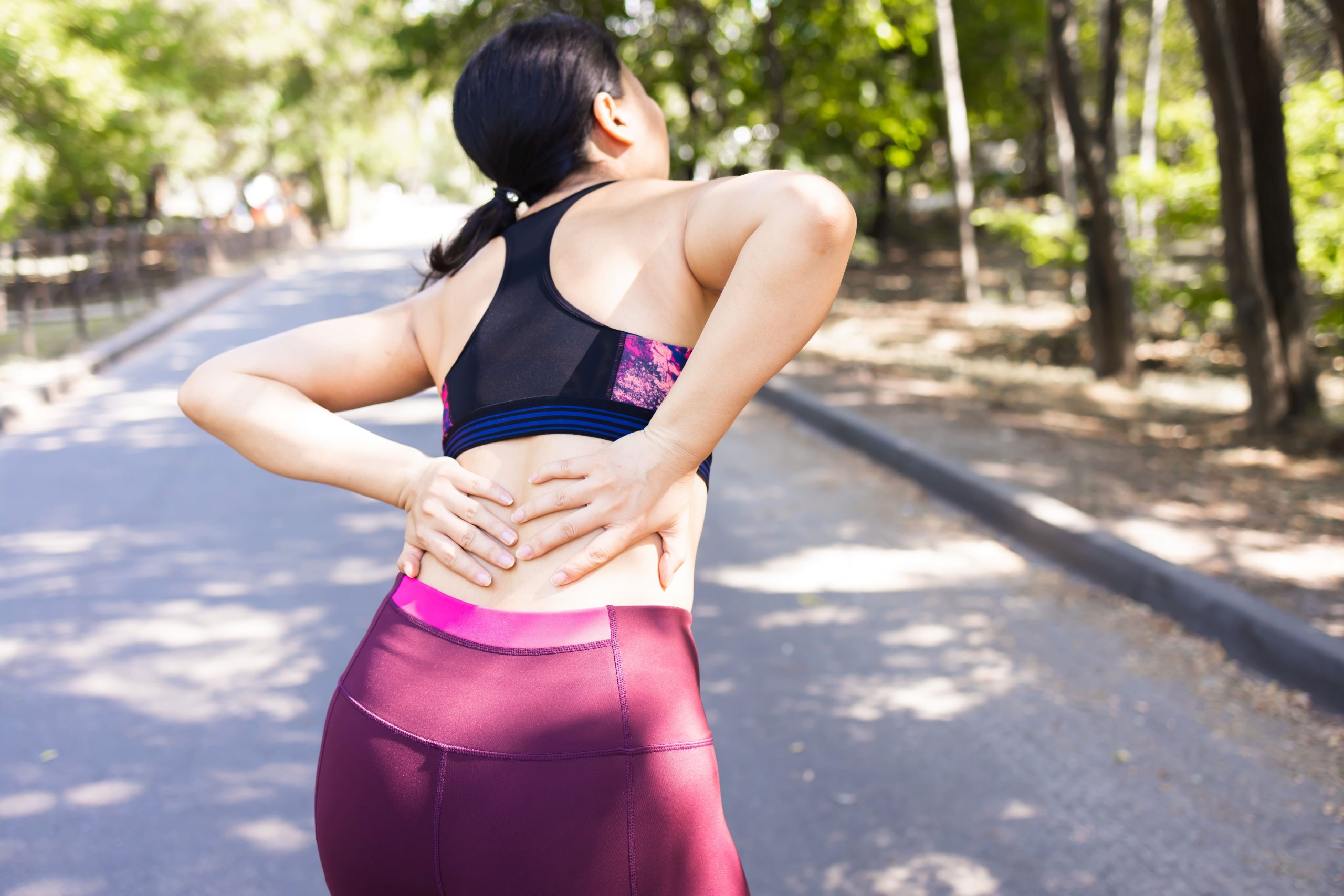
Femoroacetabular Impingement (FAI): This condition occurs when the femoral head and acetabulum rub abnormally, causing damage to the articular cartilage and labrum. Arthroscopy allows for reshaping of the bone to eliminate impingement and repair damaged tissue. FAI typically presents in younger, active individuals with groin pain exacerbated by flexion activities.
Labral Tears: The labrum is a ring of cartilage that surrounds the acetabulum, deepening the socket and enhancing joint stability. Tears may result from trauma, impingement, or degenerative changes. Arthroscopy enables surgeons to debride or repair the labrum, depending on the tear pattern and tissue quality.
Loose Bodies: Fragments of cartilage or bone can become free-floating within the joint, causing catching, locking, or pain. Arthroscopic removal of these loose bodies can provide significant symptom relief and prevent further mechanical damage to the joint surfaces.
Synovitis and Inflammatory Conditions: Inflammation of the synovial lining of the joint can cause pain and effusion. Arthroscopic synovectomy (removal of inflamed synovium) may be performed to reduce symptoms and slow disease progression in conditions such as rheumatoid arthritis or pigmented villonodular synovitis.
Benefits of Hip Arthroscopy
Hip arthroscopy offers several advantages over traditional open surgery for appropriate candidates.
Smaller Incisions: The small incisions used in arthroscopy minimise damage to surrounding muscles, tendons, and soft tissues. This reduced tissue disruption leads to less post-operative pain and a faster return to daily activities compared to open procedures.
Detailed Joint Examination: The arthroscope provides magnified views of the hip joint structures, allowing surgeons to identify and address problems directly. This helps with complex conditions that may be difficult to assess through open approaches.
Fewer Surgical Risks: Smaller incisions are associated with lower infection rates, less blood loss, and reduced risk of wound healing problems. The minimally invasive nature of the procedure also decreases the likelihood of post-operative scarring.
Same-Day Discharge: Most hip arthroscopies are performed as day cases, allowing patients to return home on the same day as surgery. This reduces hospital stay costs and allows recovery in the comfort of one’s home environment.
Patient Selection and Preoperative Assessment
The success of hip arthroscopy depends on selecting appropriate candidates. A comprehensive preoperative evaluation determines who may benefit from the procedure.
Clinical Examination: The assessment includes specific tests such as the FADIR (flexion, adduction, internal rotation) test for impingement, evaluation of range of motion, and localisation of pain. These findings help connect symptoms with specific conditions and guide treatment planning.
Imaging Studies: Plain radiographs show bony architecture, joint space, and signs of osteoarthritis. MRI arthrography reveals soft tissue structures, particularly the labrum and articular cartilage. Some cases require CT scans for complex bony abnormalities or 3D reconstruction for surgical planning.
Exclusion Criteria: Patients with advanced osteoarthritis (joint space narrowing >2mm), significant femoral head deformity, or acetabular dysplasia may not benefit from arthroscopy and may need alternative treatments such as total hip arthroplasty or periacetabular osteotomy.
Preparing for Hip Arthroscopy
Preparation before surgery helps create better outcomes and a smoother recovery process.
Medical Evaluation: A preoperative assessment including physical examination and appropriate laboratory tests confirms patients are medically fit for surgery. Existing medical conditions should be stable, and certain medications that affect bleeding may need to be temporarily stopped.
Physical Conditioning: Maintaining fitness before surgery can speed recovery. Preoperative physiotherapy focusing on hip strength and flexibility may help, particularly for patients planning to return to sports activities.
Home Adjustments: Modifying the home environment to accommodate temporary mobility limitations makes recovery easier. This includes placing needed items within reach, removing trip hazards, and arranging sleeping on the ground floor if stairs will be difficult.
Support Arrangements: Planning for assistance with transportation, household tasks, and personal care for the first few days after surgery helps the transition home. Patients cannot drive immediately after surgery and should arrange alternative transport.
Step-by-Step Procedure
Hip arthroscopy follows a systematic approach for joint access and treatment. The procedure typically includes:
Initial Setup: The procedure is performed under general anaesthesia with the patient positioned on a traction table. Controlled traction creates space between the femoral head and acetabulum, allowing for instrument access.
Portal Creation: Small incisions (portals) are made around the hip, through which the arthroscope and surgical instruments are inserted. These strategically placed portals provide access to different areas of the joint.
Joint Examination: The surgeon examines both the central compartment (inside the socket) and peripheral compartment (around the neck of the femur) to identify areas requiring treatment.
Treatment Application: Based on the specific condition, the surgeon performs the necessary interventions such as labral repair, bone reshaping, loose body removal, or cartilage treatment.
Completion: Once treatment is finished, instruments are removed, and the small incisions are closed with sutures or adhesive strips.
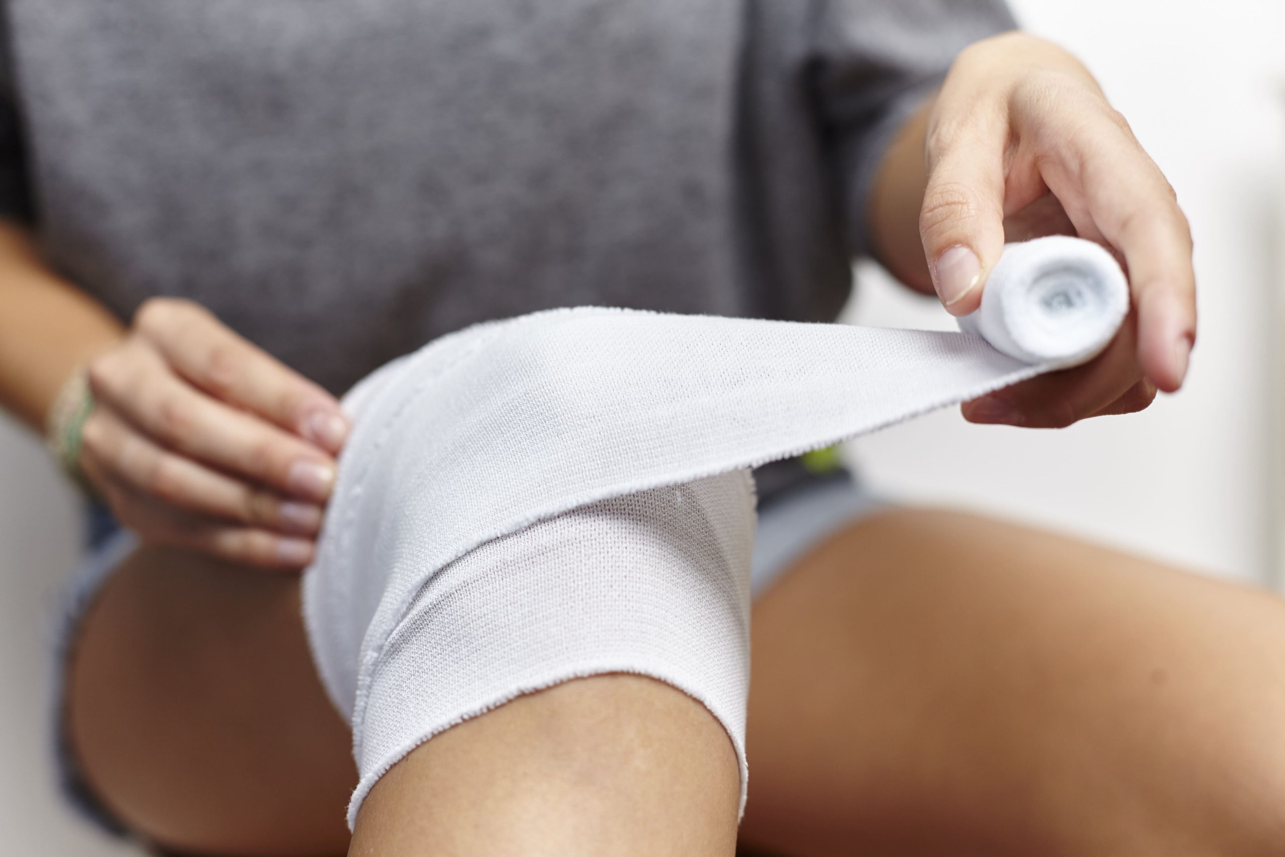
Aftercare & Recovery
The rehabilitation process following hip arthroscopy progresses through several phases:
Early Recovery (0-2 weeks): Patients use crutches with limited weight-bearing as determined by their surgeon. Pain management includes prescribed medications and ice therapy. Gentle motion exercises begin to prevent stiffness while protecting the repaired tissues. Most patients can return to desk work within two weeks.
Progressive Rehabilitation (2-12 weeks): Weight-bearing increases gradually as discomfort subsides. Physical therapy advances from basic movements to more functional exercises. Low-impact activities like stationary cycling and swimming are introduced as the hip regains strength and mobility.
Return to Activities (3-6 months): Higher-impact activities resume based on individual progress and the specific procedure performed. Athletes follow sport-specific training protocols. Most patients achieve full recovery and return to previous activity levels within 4-6 months.
Potential Complications
Hip arthroscopy carries some risks, including temporary nerve irritation from traction, numbness around incision sites, infection, and instrument-related injuries. Rare complications such as avascular necrosis or femoral neck fracture may occur. Complication rates are low, with risks decreasing when procedures are performed by experienced surgeons on appropriately selected patients.
Conclusion
Hip arthroscopy provides a minimally invasive alternative to traditional open procedures for many hip disorders. When performed for appropriate conditions in suitable patients, it delivers good outcomes with low complication rates. Success depends on proper patient selection, careful surgical execution, and dedicated rehabilitation.
Considering hip arthroscopy? Schedule a consultation today to discuss whether this procedure can help address your hip condition.