Tearing both the Anterior Cruciate Ligament (ACL) and the Medial Collateral Ligament (MCL) simultaneously compromises the knee’s stability across multiple planes, making it a fundamentally more complex injury than an isolated tear.
Typically caused by a combination of inward bending (valgus force) and rotation, these multi-ligament injuries demand specialised management strategies to address the knee’s global instability.
Because the MCL and ACL have vastly different natural healing capacities, treatment sequencing and surgical decisions must be carefully tailored to each ligament’s unique recovery profile.
Anatomy and Function of the ACL and MCL
The ACL sits deep within the knee joint. It connects the femur (thighbone) to the tibia (shinbone) in a diagonal orientation. This positioning prevents forward translation of the tibia. It also controls rotational movements during cutting, pivoting, and landing activities.
The MCL runs along the inner aspect of the knee, spanning from the femur to the tibia. Unlike the ACL, which exists within the joint capsule, the MCL lies outside the synovial environment. This anatomical difference explains their contrasting healing abilities—the MCL receives a better blood supply and often heals without surgical intervention.
How These Ligaments Work Together
During a cutting movement or lateral pivot, the ACL restrains internal rotation while the MCL prevents the knee from buckling inward. Both structures can fail simultaneously. When this happens, the knee loses stability in multiple directions. This combined instability manifests as a feeling of the knee “giving way” during activities that load the joint from different angles.
The posteromedial corner—a group of structures adjacent to the MCL—often sustains damage in combined injuries. These secondary stabilisers contribute to rotational control. Their involvement influences surgical planning and expected outcomes.
Injury Mechanisms and Classification
Combined ACL-MCL tears typically result from specific force patterns applied to the knee. Contact injuries often involve a blow to the outer knee while the foot remains planted, forcing the knee inward. Non-contact mechanisms usually combine deceleration with rotation, such as landing awkwardly from a jump or changing direction suddenly.
Grading MCL Injuries
MCL injuries follow a three-grade classification system:
- Grade I: Microscopic tearing with localised tenderness but no instability on examination
- Grade II: Partial tearing with moderate laxity when the knee is stressed in valgus
- Grade III: Complete rupture with significant gapping when stress is applied
The MCL grade significantly influences treatment decisions in combined injuries. Grade I and II MCL tears paired with ACL ruptures typically allow for a different approach than Grade III MCL tears, where both ligaments have completely failed.
Associated Injuries to Consider
Multi-ligament knee trauma rarely occurs in isolation. Meniscal tears (tears in the knee cartilage) frequently accompany combined ACL-MCL injuries, particularly involving the medial meniscus. Bone bruising on MRI indicates areas of cartilage compression during the injury event. The posterolateral corner and posterior cruciate ligament require assessment to rule out more extensive multi-ligament patterns.
Diagnostic Approach
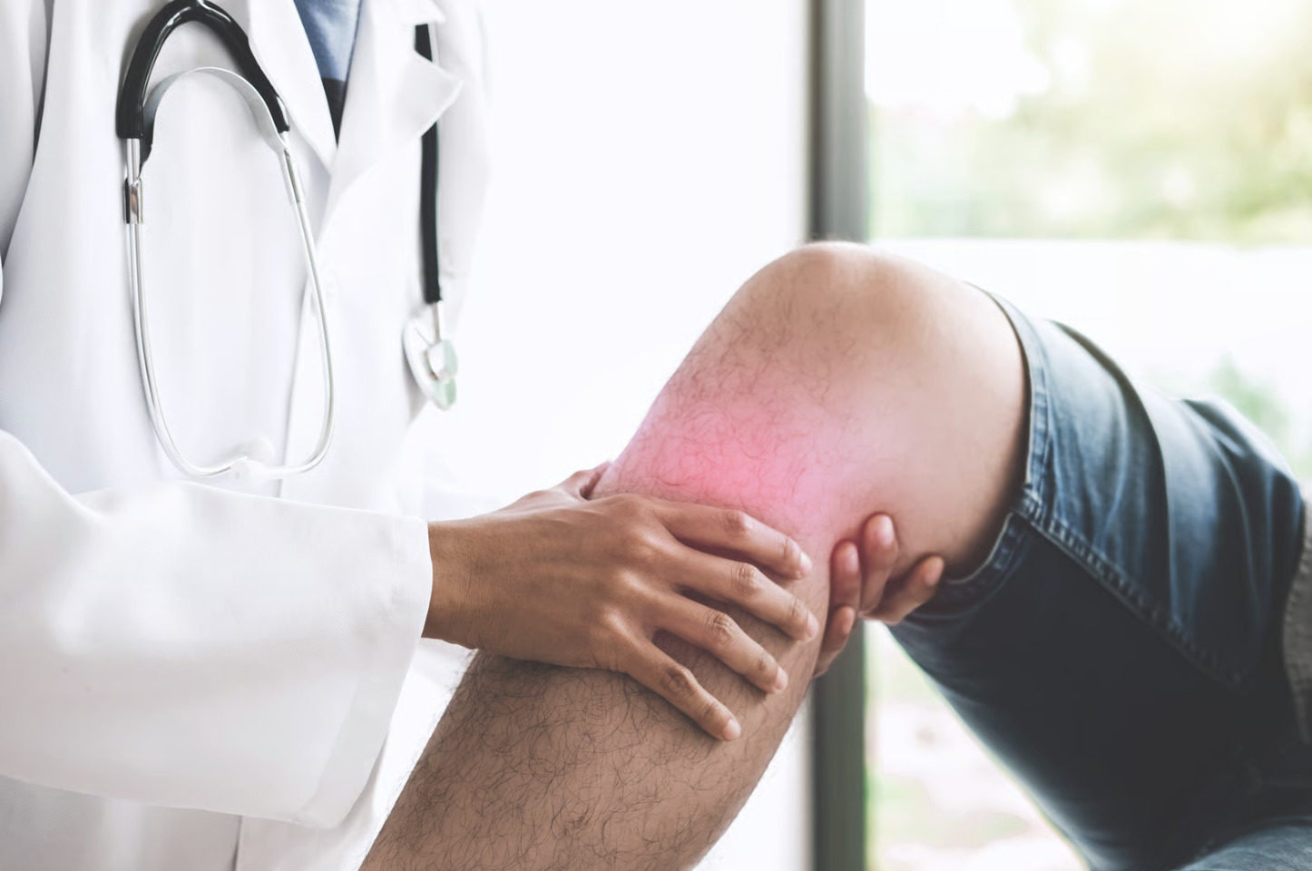
Clinical examination provides useful information before imaging (such as X-rays or MRIs). Swelling patterns differ between isolated and combined injuries—significant medial-sided swelling alongside generalised knee effusion suggests MCL involvement in addition to intra-articular pathology.
Physical Examination Tests
The Lachman and anterior drawer tests assess ACL integrity by measuring tibial translation, while the pivot shift test reproduces the specific instability caused by a deficiency in this ligament. To evaluate the MCL, surgeons perform valgus stress testing at different angles; instability at full extension often indicates more extensive damage to the knee’s medial-sided structures.
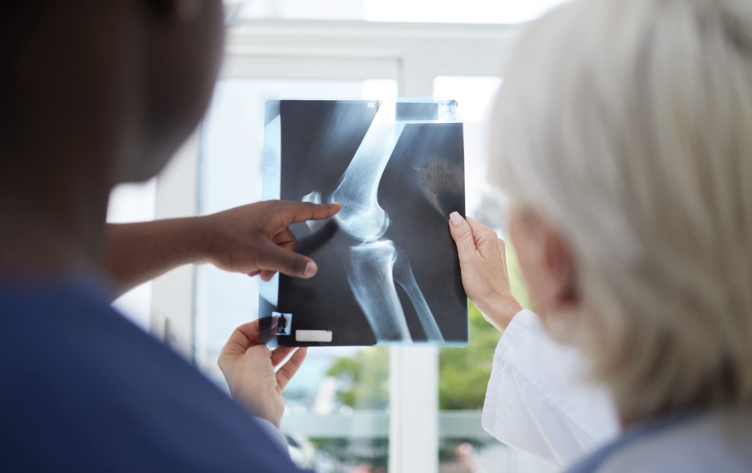
Imaging Studies
MRI provides high-resolution views of soft tissues, allowing doctors to identify the exact location and severity of ACL and MCL tears by abnormal signal intensities. In cases of significant Grade III laxity, stress radiographs may be used to quantify joint opening and assist in precise surgical planning.
Treatment Decision-Making
The management of combined ACL-MCL injuries is highly personalised, with surgeons weighing the MCL grade and the patient’s activity goals against the timing of the trauma. By tailoring the approach to specific injury patterns, doctors can decide whether a staged or simultaneous treatment plan will yield the functional outcome.
Non-Operative MCL Management
Because the MCL is located outside the joint capsule and has a robust blood supply, Grade I and II tears often heal effectively through protected weight-bearing and functional bracing. This natural healing capacity allows for a staged treatment strategy, where the medial side of the knee is stabilised non-surgically before proceeding with ACL reconstruction.
Surgical Considerations
Timing is critical in surgical planning; operating too soon can lead to excessive scarring, while waiting too long may result in muscle atrophy or further joint wear. While lower-grade MCL tears are typically allowed to heal first, Grade III ruptures often require a more complex decision between concurrent reconstruction of both ligaments or a staged surgical approach.
Surgical Techniques
ACL reconstruction uses graft tissue to replace the torn ligament. Graft options include:
- Hamstring tendons: Harvested from the same knee, these grafts cause less anterior knee pain but may slightly weaken knee flexion strength
- Patellar tendon: Provides bone-to-bone healing at fixation points but carries the risk of anterior knee discomfort
- Quadriceps tendon: Used by some surgeons, offering good graft properties with acceptable donor site effects
- Allograft (donor tissue): Avoids harvest site complications but may have slightly different incorporation characteristics
MCL reconstruction, when necessary, uses similar graft options to recreate the ligament’s anatomical course along the medial knee.
Rehabilitation Phases
Recovery from combined ligament injuries extends longer than single-ligament rehabilitation. It typically spans nine to twelve months before return to unrestricted activity.
Early Phase
The primary focus is on protecting the healing structures while re-establishing quadriceps activation and preventing joint stiffness through immediate range-of-motion exercises. Patients work toward achieving full knee extension and gradual flexion, using techniques such as patellar mobilisation to prevent the formation of limiting scar tissue.
Intermediate Phase
As the tissue stabilises, rehabilitation intensifies with closed-chain exercises like squats and leg presses to build strength through controlled joint compression. This phase also introduces proprioceptive training to retrain the knee’s sensors, restoring the body’s awareness of joint position and movement.
Advanced Phase
Running progression usually begins with straight-line jogging at the three-to-four-month mark, eventually advancing to more complex direction changes. Progression is strictly milestone-based, requiring adequate quadriceps strength and the absence of swelling before introducing sport-specific drills.
Return to Sport Phase
The final stage focuses on explosive power, agility, and objective return-to-sport testing, such as hop tests, to ensure the knee is physically resilient. Because psychological readiness often lags behind physical healing, addressing the fear of re-injury is a vital component of a successful return to competition.
Factors Affecting Outcomes
Several variables influence recovery trajectory and long-term knee function after combined ligament injuries. Results depend on your unique health status and injury characteristics.
Patient-Related Factors
Age affects healing capacity and tissue quality, though motivated patients across age groups can achieve good functional outcomes. Pre-injury activity level and muscle conditioning influence the starting points of rehabilitation. Smoking impairs tissue healing and increases the risk.
Injury-Related Factors
The number of damaged structures correlates with the complexity of the outcome. Meniscal tears requiring repair add rehabilitation restrictions and extend timelines. Cartilage damage at the time of injury affects long-term joint health regardless of ligament reconstruction success.
Treatment-Related Factors
Graft choice, surgical technique, and fixation method all influence early rehabilitation and eventual outcomes. Compliance with rehabilitation protocols strongly affects final results; patients who complete structured physiotherapy programmes demonstrate better strength recovery and functional scores.
Long-Term Considerations
Reconstructed knees function well for daily activities and many recreational sports. However, multi-ligament injuries carry higher rates of post-traumatic arthritis (joint degeneration that develops after an injury) compared to isolated tears, even with successful reconstruction. This occurs because the initial injury often damages cartilage. Altered joint mechanics—even subtle changes—can accelerate wear over time.
Activity modification may become necessary as the knee ages. Many patients transition from high-demand pivoting sports to lower-stress activities while maintaining fitness and quality of life.
Managing Your Recovery Effectively
- Commit to physiotherapy: Consistent attendance and home exercise completion correlate directly with outcome quality
- Progress based on milestones: Advancing when criteria are met rather than at arbitrary time points reduces setback risk
- Communicate symptoms: Increasing pain, swelling, or instability during rehabilitation requires assessment and potential programme modification
- Address psychological barriers: Fear of movement and re-injury are normal—discuss these concerns with your treatment team
- Plan for the long term: Building habits around knee-friendly exercise and maintaining strength provides ongoing joint protection
When to Seek Professional Help
- Knee giving way during walking or daily activities
- Significant swelling following a twisting or contact injury
- Inability to fully straighten or bend the knee after trauma
- Pain along the inner knee combined with a sense of instability
- Persistent symptoms despite initial rest and conservative measures
- Progressive difficulty with stairs or uneven surfaces
Commonly Asked Questions
How long before I can return to sports after combined ACL-MCL surgery?
Return to pivoting sports typically requires nine to twelve months of rehabilitation, though the timeline and degree of readiness vary from person to person. Progression is based on achieving strength, movement, and psychological readiness criteria. Some patients return to linear activities earlier, such as jogging or cycling.
Will I need to wear a brace permanently?
Most patients discontinue bracing after completing rehabilitation. Some choose to wear a functional brace during higher-risk activities for added confidence, though evidence for injury prevention in reconstructed knees remains limited.
Can I avoid surgery if both ligaments are torn?
Isolated MCL tears often heal without surgery. However, ACL tears in active individuals typically require reconstruction to restore stability for pivoting activities. Non-operative management may suit patients with lower activity demands who can modify their activities to avoid instability episodes.
What happens if I delay treatment?
Prolonged instability can cause additional meniscal and cartilage damage from repeated giving-way episodes. However, surgery performed months after injury can still achieve good outcomes—the knee doesn’t have a strict expiration date for reconstruction.
Is the reconstructed knee as strong as the original?
Graft tissue undergoes remodelling over twelve to eighteen months. It eventually achieves properties similar to native ligament. Functional outcomes in well-rehabilitated patients often match or approach pre-injury levels, though subtle differences in proprioception may persist.
Next Steps
In combined ACL-MCL injuries, treatment sequencing matters: allowing the MCL to heal before ACL reconstruction takes advantage of the MCL’s natural repair capacity and creates more stable surgical conditions. Returning to pivoting sports requires meeting objective strength and movement criteria—not simply reaching a time milestone—and typically spans 9 to 12 months. Meniscal or cartilage involvement at the time of injury warrants discussion with your surgeon, as it affects both rehabilitation timelines and long-term joint health.
If you are experiencing knee instability, giving-way episodes, or inner knee pain following a twisting or contact injury, consult an orthopaedic knee surgeon specialising in knee ligament injuries for a comprehensive evaluation and discussion of treatment options.

Experiencing Knee Pain or Injury?
Get a Personalised Treatment Plan
Find relief with our hip & knee specialists.
Make An Enquiry