Can a piece of bone in your knee actually die and break away from the joint? In osteochondritis dissecans (OCD), this exact process occurs when bone beneath the cartilage loses its blood supply, causing both bone and cartilage to separate from surrounding tissue. The knee joint—particularly the medial femoral condyle, the rounded end of the thighbone on the inner side, is the most commonly affected location. This condition predominantly affects adolescents and young adults during skeletal growth, though it can occur at any age. The separated fragment may remain partially attached, become completely loose within the joint space, or heal spontaneously, depending on the patient’s skeletal maturity and lesion stability.
How OCD Develops in the Knee
The exact mechanism triggering osteochondritis dissecans remains under investigation. However, repetitive microtrauma (small, repeated injuries) appears to play a significant role. Young athletes participating in high-impact sports—particularly those involving running, jumping, and pivoting—place repeated stress on the knee joint surfaces. Over time, this cumulative loading may disrupt blood flow to a localised area of subchondral bone (the bone layer directly beneath the cartilage).
Vascular insufficiency (reduced blood flow) prevents normal bone maintenance and repair. The affected bone segment gradually weakens. It may begin separating from healthy surrounding tissue. The overlying articular cartilage (the smooth, protective layer covering the joint surface) depends on the underlying bone for structural support and nutrition. Subsequently, it deteriorates.
Genetic predisposition may contribute to susceptibility. Some individuals develop OCD in multiple joints or have family members with similar conditions. Skeletal variants that alter joint mechanics or loading patterns may also increase risk.
Recognising Signs and Symptoms
Early-stage OCD often produces vague, activity-related discomfort. Pain typically localises to the front or inner aspect of the knee. It worsens with weight-bearing activities, stair climbing, or prolonged walking. Rest usually provides relief during the initial stages.
As the lesion progresses, symptoms become more pronounced and specific:
- Swelling develops within the joint, particularly after activity
- Stiffness limits full knee flexion (bending) or extension (straightening)
- Catching or locking sensations occur if loose fragments interfere with joint movement
- Giving way episodes happen when the knee feels unstable during weight-bearing
- Audible clicking may accompany knee movement
Patients with unstable or detached fragments often report a sudden onset of severe symptoms. These include the inability to fully straighten the knee due to mechanical blocking by loose bodies within the joint.
Diagnostic Evaluation Process
Clinical examination begins with assessment of gait patterns, knee alignment, and range of motion. Palpation (gentle pressing) may reveal tenderness over the affected condyle. The Wilson test—internal rotation of the tibia (shin bone) during knee extension—can reproduce pain by pressing the tibial spine against the typical lesion location.
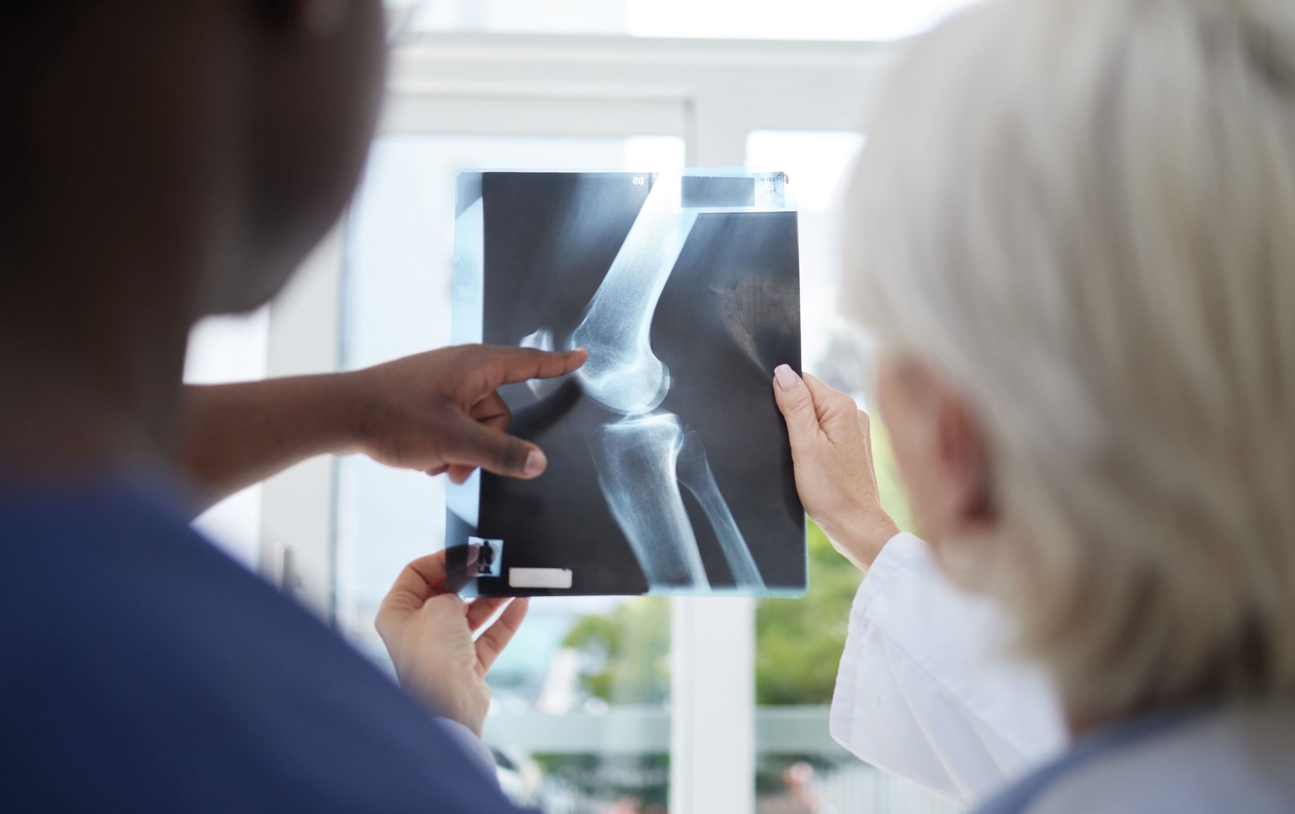
Plain radiographs (X-rays) provide initial imaging evaluation. Standard anteroposterior, lateral, and tunnel (notch) views may reveal the characteristic radiolucent lesion appearing as a demarcated fragment of subchondral bone. Tunnel views prove particularly valuable for visualising the posterior aspect of the femoral condyles, where many lesions occur.
Magnetic resonance imaging (MRI), a scan that uses magnetic fields to create detailed images of soft tissues and bone, offers a detailed assessment of lesion characteristics for treatment planning:
- Lesion size measured in three dimensions
- Cartilage surface integrity indicates whether the fragment remains covered
- Fragment stability was assessed by the fluid signal between the fragment and the parent bone
- Bone marrow oedema suggesting ongoing disease activity
MRI findings directly influence treatment decisions, particularly regarding surgical versus non-surgical management.
💡 Did You Know?
The knee’s medial femoral condyle bears significant load during normal walking—forces much higher than body weight pass through this area with each step, explaining why this location frequently develops OCD lesions.
Classification and Staging
Orthopaedic specialists (doctors who specialise in treating bone, joint, and muscle conditions) classify OCD lesions based on stability, skeletal maturity, and imaging characteristics. This classification guides treatment selection and prognosis estimation.
Juvenile OCD occurs in patients with open growth plates (physes, areas of developing cartilage near the ends of bones) visible on radiographs. These patients generally have better healing potential due to robust blood supply and growth capacity in immature bone. The presence of open physes indicates significant remaining skeletal growth.
Adult OCD affects patients with closed growth plates and fully mature skeletons. Healing capacity diminishes after skeletal maturity. Adult lesions more frequently require surgical intervention.
Lesion stability assessment categorises fragments as:
- Stable: Intact cartilage surface, no fluid signal behind fragment on MRI, fragment remains attached
- Unstable: Cartilage breach, fluid completely surrounding fragment, partial or complete detachment
Non-Surgical Treatment Approaches
Conservative management is appropriate for stable lesions in skeletally immature patients. A healthcare professional can discuss personalised treatment goals based on individual factors, including age, lesion characteristics, activity level, and overall health. The treatment protocol centres on activity modification and protected weight-bearing to reduce mechanical stress on the healing bone.
Activity restriction eliminates high-impact sports, running, and jumping for extended periods—typically for three to six months. Low-impact activities, such as swimming and cycling, may continue if they remain pain-free.
Protected weight-bearing using crutches reduces compressive forces through the affected condyle. Complete non-weight-bearing may be necessary for larger or more symptomatic lesions. Gradual weight-bearing progression follows clinical and radiographic improvement.
Bracing with a hinged knee brace provides stability. It may limit the range of motion to positions that minimise lesion loading. Brace wear continues throughout the healing period.
Physical therapy maintains quadriceps and hamstring strength, preserves range of motion, and addresses any gait abnormalities. Therapy intensity increases as healing progresses.
Serial imaging—typically radiographs every six to twelve weeks—monitors lesion healing. MRI reassessment is performed if radiographic changes occur or symptoms persist. Healing evidence includes lesion reintegration, decreased surrounding oedema, and resolved radiolucency.
Surgical Treatment Options
Surgery becomes necessary when conservative treatment fails, lesions demonstrate instability, fragments detach, or adult patients present with symptomatic OCD. Several surgical techniques address different lesion presentations.
Arthroscopic Drilling
For stable lesions with intact cartilage surfaces, the surgeon creates channels through the overlying cartilage or through healthy bone adjacent to the lesion. This allows blood vessels to grow into the affected area. It promotes healing of the bone segment that has lost its blood supply. Multiple small holes encourage a new blood supply without destabilising the fragment.
Fragment Fixation
Unstable but salvageable fragments benefit from internal fixation using bioabsorbable pins, screws, or bone pegs. The surgeon repositions the fragment to its original location. They then secure it in place, allowing for biological healing while maintaining the congruity of the cartilage surface. Hardware selection depends on fragment size, bone quality, and surgeon preference.
Fragment Excision and Marrow Stimulation
Unsalvageable fragments—those too damaged, fragmented, or calcified for repair—require removal. After removing the fragment, the surgeon performs marrow stimulation techniques such as microfracture. Small perforations in the underlying bone release stem cells (specialised cells that can develop into various tissue types) and growth factors. These generate fibrocartilage (a type of repair cartilage) to fill the defect.
Osteochondral Grafting
Large defects or failed primary procedures may necessitate osteochondral transplantation (transfer of bone and cartilage tissue). Autograft procedures involve the surgeon harvesting plugs of bone and cartilage from non-weight-bearing areas of the patient’s own knee for transplantation into the defect. Allograft tissue from donors provides an alternative for extensive defects.
⚠️ Important Note
Post-surgical rehabilitation requires strict adherence to weight-bearing restrictions and activity limitations. Premature loading can displace fixed fragments or damage healing repair tissue.
Recovery and Rehabilitation Timeline
Recovery duration varies considerably based on treatment type, lesion characteristics, and individual healing response.
Post-drilling recovery typically involves six weeks of protected weight-bearing followed by gradual activity progression. Return to sports occurs around four to six months after surgery. This depends on clinical and imaging evidence of healing.
Post-fixation rehabilitation requires longer protected weight-bearing—often six to twelve weeks—while the fixed fragment incorporates. Range-of-motion exercises begin early, but loading progresses slowly. Sports return generally occurs six to nine months postoperatively.
Post-microfracture protocols emphasise prolonged non-weight-bearing (six to eight weeks) to protect the developing repair tissue. Continuous passive motion machines may be used to nourish the healing cartilage. Full activity return takes nine to twelve months.
Physical therapy progresses through phases:
- Range of motion restoration
- Strength rebuilding
- Functional training
- Sport-specific preparation
Each phase must be completed before advancing to prevent setbacks.
Long-Term Outlook and Joint Health
Treatment of OCD aims to preserve native cartilage, restore joint surface congruity, and prevent premature degenerative changes. Outcomes differ among patients based on several factors. These include patient age at treatment, lesion size and location, treatment type, and cartilage quality at the time of intervention.
Juvenile patients with stable lesions treated conservatively can achieve complete healing and return to full activity. Adult patients and those requiring surgery for unstable lesions face higher rates of residual symptoms and subsequent degenerative changes.
Patients with a history of knee OCD benefit from:
- Activity modification, avoiding repetitive high-impact loading
- Weight management, reducing joint stress
- Regular low-impact exercise maintains muscle strength and joint mobility
- Prompt attention to new knee symptoms
When to Seek Professional Help
- Persistent knee pain lasting more than several weeks despite rest
- Swelling that recurs with activity or appears spontaneously
- Mechanical symptoms (such as catching, locking, or giving way)
- Inability to fully bend or straighten the knee
- Pain that interferes with daily activities or sleep
- Previous OCD diagnosis with new or worsening symptoms
Commonly Asked Questions
Can osteochondritis dissecans heal without surgery?
Stable lesions in skeletally immature patients frequently heal with non-surgical treatment. This includes activity modification and protected weight-bearing. Adult patients and those with unstable lesions often require surgery.
How long will recovery take?
Recovery timelines range from three to twelve months. The duration depends on treatment type and individual healing. Conservative treatment for stable lesions typically requires four to six months of activity restriction. Surgical recovery varies by procedure complexity. More extensive interventions require longer rehabilitation.
Will I develop arthritis after OCD?
OCD increases the risk of osteoarthritis (degenerative joint disease, in which cartilage breaks down over time), particularly when significant cartilage damage occurs or when treatment is delayed. Treatment that preserves cartilage and restores smoothness to the joint surface may help reduce the risk of future degenerative changes, though individual outcomes vary with lesion severity, treatment timing, and personal health factors.
Can I return to sports after treatment?
Many patients return to full athletic activity following treatment, though the timeline and degree of return vary from person to person. Return timing depends on lesion healing confirmed by imaging, restoration of strength and range of motion, and completion of sport-specific rehabilitation. High-impact sports carry a higher risk of re-injury or new lesion development.
Does OCD affect both knees?
Bilateral involvement occurs in some OCD cases. Orthopaedic evaluation typically includes imaging of both knees when OCD appears in one knee, particularly in younger patients.
Next Steps
Early intervention for stable OCD lesions offers the opportunity for healing and joint preservation. Accurate diagnosis through orthopaedic evaluation determines lesion stability, appropriate treatment selection, and long-term prognosis.
If you’re experiencing persistent knee pain, swelling, catching, or locking, schedule a consultation with an orthopaedic knee surgeon for comprehensive evaluation and treatment planning.

Experiencing Knee Pain or Injury?
Get a Personalised Treatment Plan
Find relief with our hip & knee surgeons.
Make An Enquiry