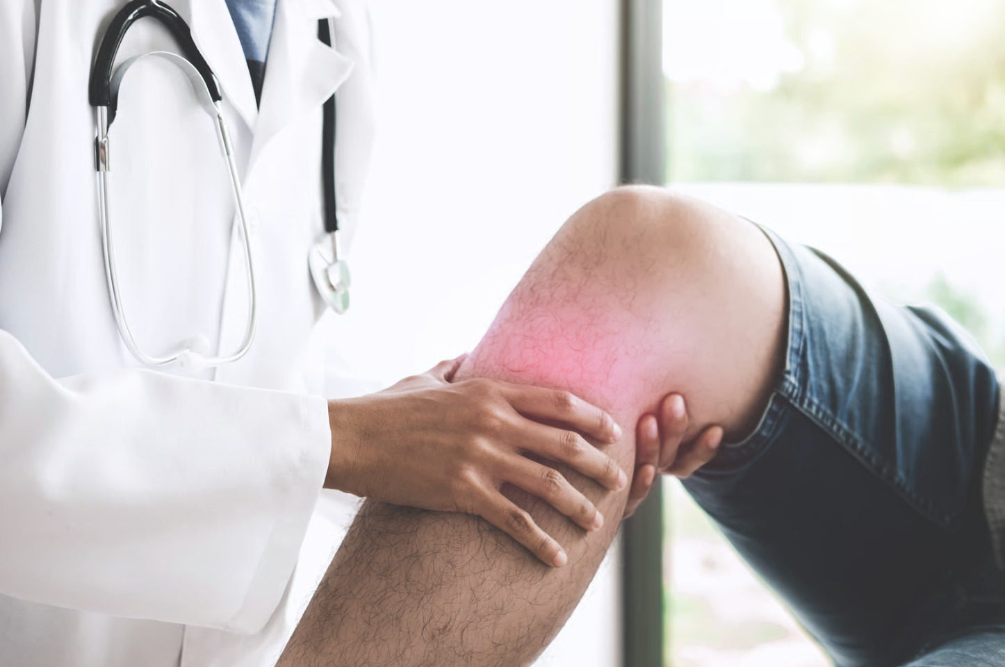
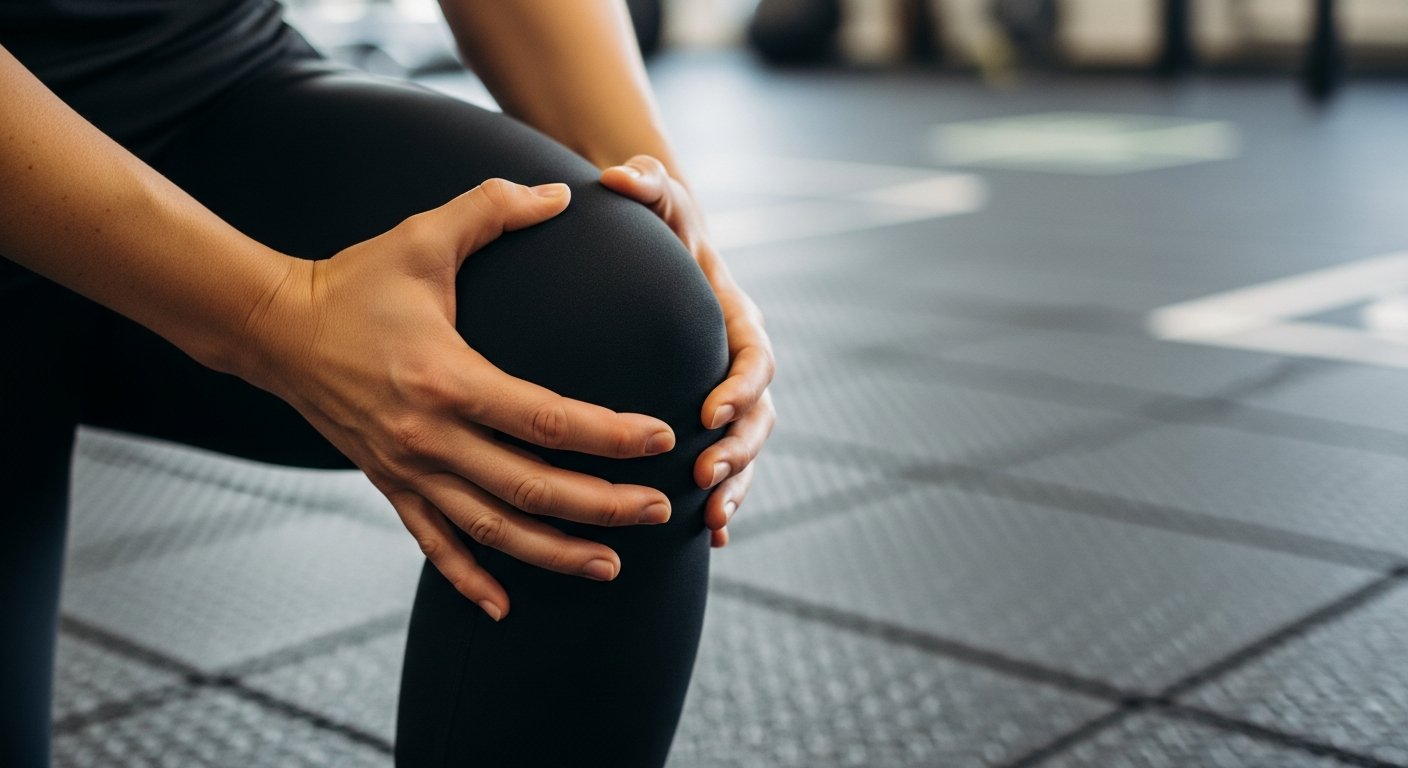
Does your knee hurt more when you squat deeper? Knee pain during or after squatting typically originates from increased compression forces across the patellofemoral joint, which is the area where your kneecap meets your thighbone. These forces can exceed several times your body weight during deep flexion. The squatting motion places significant demands on cartilage, ligaments, and the menisci—especially when the knee bends beyond a certain angle. Pain location provides valuable diagnostic information because anterior knee discomfort often suggests patellofemoral involvement, while pain along the joint line may indicate meniscal pathology.
Patellofemoral Pain Syndrome
Patellofemoral pain syndrome is a leading cause of discomfort at the front of the knee during squatting, occurring when the kneecap tracks improperly within its groove on the thighbone. This misalignment causes an uneven pressure distribution across the cartilage surface, which becomes more pronounced as the knee bends and compressive forces reach multiple times your body weight.
-
Symptom Progression: Individuals typically experience a dull, aching pain around the kneecap that intensifies during stair climbing, squatting, or prolonged sitting. Some may also notice grinding or clicking sensations as the knee moves through its range of motion.
-
Muscular Weakness: Weakness in the quadriceps, specifically the vastus medialis obliquus at the front of the thigh, prevents the kneecap from staying stabilised. This is often paired with weak hip abductors, which are the muscles responsible for moving the leg away from the body.
-
Biomechanical Stress: When these muscle imbalances or anatomical variations exist, the thigh rotates inward excessively during weight-bearing activities. This forces certain areas of the cartilage to bear disproportionate stress rather than distributing the load evenly.
Meniscal Injuries
The menisci—crescent-shaped cartilage structures that cushion the space between your thighbone and shinbone—face considerable stress during deep squatting. These structures distribute load across the knee joint and provide stability during rotational movements.
Meniscal tears occur through two primary mechanisms:
- Acute tears typically result from sudden twisting or pivoting movements while the foot remains planted.
- Degenerative tears develop gradually as meniscal tissue becomes less resilient with age, sometimes tearing during routine activities such as deep squatting or kneeling.
Pain from meniscal pathology often localises to the inner or outer joint line, depending on which meniscus is affected. Many individuals report mechanical symptoms (such as catching, locking, or the knee giving way). Swelling may develop within hours of an acute injury or intermittently with degenerative tears.
The posterior horn of the medial meniscus (the back portion of the inner shock absorber) bears substantial load during deep knee flexion. This explains why squatting frequently aggravates tears in this location. Pain that specifically worsens at end-range flexion suggests possible posterior horn involvement.
Osteoarthritis
Osteoarthritis develops when articular cartilage—the smooth covering on bone surfaces within joints—progressively wears down. The knee contains three compartments where this degeneration can occur:
- The medial (inner) compartment
- The lateral (outer) compartment
- The patellofemoral compartment
Squatting-related pain in osteoarthritis typically correlates with the compartment most involved. Patellofemoral arthritis causes anterior knee pain during squatting. Tibiofemoral arthritis (arthritis affecting the main weight-bearing surfaces between the thighbone and shinbone) may cause broader discomfort during the loaded portions of the movement.
Morning stiffness that is transient characterises osteoarthritis, distinguishing it from inflammatory conditions. Many individuals notice worsening symptoms after periods of increased activity, with some improvement following rest. Weather changes, particularly declines in barometric pressure, appear to influence symptom severity in some patients.
Radiographic findings don’t always correlate with symptom severity. Some individuals with significant visible changes experience minimal discomfort, while others with modest findings report substantial pain.
Patellar Tendinopathy
Patellar tendinopathy affects the tendon connecting the kneecap to the shinbone. It develops from repetitive loading that exceeds the tissue’s capacity to recover.
Pain localises specifically to the inferior pole of the patella—the lower tip of the kneecap where the tendon attaches. Unlike patellofemoral syndrome, which produces diffuse anterior knee pain, tendinopathy causes well-localised tenderness that patients can often pinpoint with one finger.
The tendon typically tolerates activities at lower loads but becomes symptomatic when demands increase. Deep squatting, particularly with added weight, often reproduces symptoms. Pain typically presents at the beginning of activity, may improve with warm-up, and then returns after exercise cessation.
Tendon structure changes accompany this condition, with disorganisation of collagen fibres and increased vascularity visible on ultrasound or MRI. These structural changes explain why rest alone rarely resolves tendinopathy; active rehabilitation that progressively loads the tendon is a key treatment approach.
Iliotibial Band Syndrome
The iliotibial band is a thick fibrous structure running along the outer thigh that often contributes to lateral knee pain during squatting. This band crosses a bony prominence on the outer knee, the lateral femoral epicondyle, where repetitive friction at the interface can cause significant irritation.
-
Biomechanical Stress: Squatting with poor form can irritate this structure, particularly when the knees cave inward during the movement. This misalignment increases tension along the outer knee and aggravates the band against the underlying bone.
-
Symptom Presentation: Pain is typically localised on the lateral aspect of the knee and is often accompanied by tenderness over the bony prominence. Symptoms usually worsen as the knee moves through the specific angle of flexion where maximum friction occurs.
-
Proximal Weakness: Hip abductor weakness contributes significantly to this condition by permitting excessive inward movement of the thigh during weight-bearing. Strengthening these muscles, which move the leg away from the body, is often just as important as local treatment.
Biomechanical Factors That Increase Risk
Squatting technique significantly influences the distribution of forces across knee structures. Several modifiable factors contribute to the development of knee pain during this movement pattern.
Foot and Ankle Mobility
Limited ankle dorsiflexion (the movement that brings your toes toward your shin) restricts the extent to which the knee can travel forward during squatting. To compensate, individuals often shift weight backward, increasing demand on the posterior chain, or allow the heels to lift—which shifts load anteriorly onto the patellofemoral joint. Alternatively, excessive foot pronation (flattening) can lead to internal rotation of the lower leg, potentially affecting patellar tracking.
Hip Mobility and Control
Restricted hip flexion results in lumbar spine and knee compensation through excessive motion. Similarly, inadequate hip external rotation contributes to knee valgus (inward collapse) during squatting—a position that increases lateral patellofemoral stress and iliotibial band tension.
Muscle Imbalances
Quadriceps dominance relative to hip extensor strength shifts squatting mechanics in ways that increase knee loading. When the gluteal muscles (buttocks) contribute insufficiently to the movement, the quadriceps must generate greater force, thereby increasing patellofemoral compression.
💡 Did You Know?
The articular cartilage in your knee lacks blood vessels and relies on joint fluid for nutrition. Gentle movement helps distribute this fluid across cartilage surfaces, which is why complete rest often proves less beneficial than modified activity for many knee conditions.
Modifying Squatting Technique
Several adjustments can reduce knee stress while maintaining training benefits for those experiencing squatting-related pain. Modifying your technique allows for continued strength development while managing the load placed on sensitive joint structures.
-
Box Squats and Tempo: Box squats limit depth and provide a target for consistent positioning, helping control the descent and preventing rapid loading. Adding tempo modifications, such as slowing the lowering phase, reduces momentum and peak forces while building tissue resilience over time.
-
Stance Width Alterations: Changing your stance width shifts which structures bear the load. Wider stances typically reduce patellofemoral stress by increasing the demand on the hip muscles, whereas narrower stances tend to do the opposite.
-
Heel Elevation: Using weightlifting shoes or small plates under the heels reduces the requirement for ankle dorsiflexion and shifts loading toward the quadriceps. This modification helps some individuals with posterior knee pain, though it may aggravate patellofemoral symptoms in others.
When to Seek Professional Help
- Knee pain persists beyond several weeks despite activity modification
- Swelling that develops rapidly after injury or recurs with activity
- Mechanical symptoms (such as locking, catching, or the knee giving way)
- Pain that wakes you from sleep
- Visible deformity or inability to fully straighten the knee
- Progressive worsening despite rest and conservative measures
- Pain accompanied by warmth or redness around the joint
- Instability or feeling that the knee might buckle during daily activities
Clinical Evaluation Process
Orthopaedic assessment of squatting-related knee pain typically begins with a detailed history. This includes understanding exactly when symptoms occur, what aggravates and relieves them, and how they’ve evolved over time.
Physical examination includes assessing alignment, observing gait patterns, and testing the range of motion. Specific provocative tests help identify involved structures: the McMurray test for meniscal pathology, patellar compression and tracking assessments for patellofemoral dysfunction, and various ligamentous stress tests when instability symptoms exist.
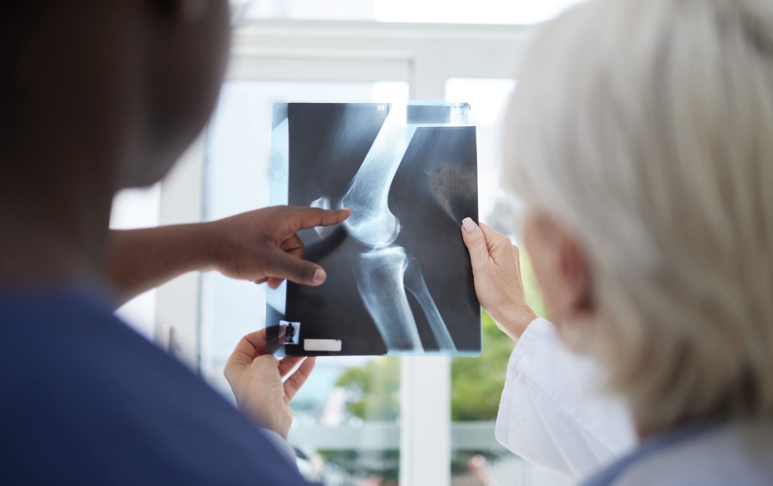
Imaging may include weight-bearing radiographs (X-rays obtained with the patient standing) to assess joint alignment and arthritic changes. MRI provides detailed soft tissue visualisation when meniscal tears, ligament injuries, or subtle cartilage damage require evaluation. Ultrasound offers a dynamic assessment of tendons and can guide injections when indicated.
Treatment Approaches
Management varies substantially based on diagnosis, with many common conditions initially responding to non-surgical interventions. A healthcare professional can discuss treatment options tailored to your specific diagnosis, symptom severity, activity demands, and overall health profile.
Physiotherapy addresses modifiable factors, including muscle weakness, limited range of motion, and movement pattern abnormalities. Progressive loading programmes are used for tendinopathy and patellofemoral conditions.
Activity modification—not necessarily complete rest—allows tissue recovery while maintaining fitness. Cross-training with lower-impact activities such as cycling or swimming often permits continued exercise without exacerbating symptoms.
Bracing or taping may provide symptomatic relief for patellofemoral pain, though evidence for long-term benefit remains limited. These interventions appear to work by altering sensory feedback rather than by mechanical correction.
Injection therapies, including corticosteroids (anti-inflammatory medications) and hyaluronic acid preparations (lubricants), can provide symptom relief for certain conditions. Appropriate patient selection determines effectiveness; corticosteroids generally provide short-term benefit but may affect tissue integrity with repeated use.
Surgical intervention may be considered when conservative measures fail to adequately address symptoms. Arthroscopic procedures (minimally invasive surgery using a small camera and instruments) can address meniscal tears, remove loose bodies, or perform cartilage procedures. Realignment surgeries help specific patellofemoral conditions. Joint replacement provides definitive treatment for advanced arthritis refractory to other interventions.
Commonly Asked Questions
Can I continue squatting if I have knee pain?
This depends entirely on the underlying cause and the severity of the symptoms. Many conditions improve with modified squatting—adjusting depth, load, or technique—rather than complete avoidance. However, pushing through significant pain risks worsening tissue damage. If simple modifications don’t reduce symptoms, clinical evaluation helps determine appropriate activity levels.
How long does knee pain from squatting typically take to resolve?
Recovery timelines vary considerably. Mild muscle strain or overuse may settle within a short period with appropriate rest. Patellofemoral syndrome often requires several months of targeted rehabilitation. Meniscal tears and tendinopathy may need even longer to adequately rehabilitate, with some cases requiring surgical intervention for resolution.
Are deep squats harmful to the knees?
Deep squats aren’t inherently harmful for healthy knees with adequate mobility and strength. However, they do increase loading across certain structures—particularly the patellofemoral joint and posterior meniscal horns. Individuals with existing pathology in these areas may benefit from limiting depth. Technique and individual anatomy matter more than absolute depth.
Should I use a knee sleeve or brace when squatting?
Knee sleeves provide compression and warmth, which some individuals find comfortable during activity. They don’t prevent injury in healthy knees but may offer symptomatic relief for minor discomfort. Specific braces designed for patellofemoral conditions can alter tracking mechanics. Whether these interventions help you depends on your underlying condition.
When would surgery be considered for knee pain from squatting?
Surgical intervention typically follows failed conservative treatment lasting several months. Specific indications include displaced meniscal tears causing mechanical symptoms, loose bodies within the joint, or significant cartilage damage amenable to repair or restoration procedures. The decision incorporates symptom severity, imaging findings, activity demands, and response to non-operative care.
Next Steps
Determine whether your pain stems from patellofemoral dysfunction, meniscal involvement, or tendinopathy through clinical evaluation. Modify your squatting depth, stance width, or loading based on your specific condition. Address contributing factors such as ankle mobility restrictions or hip weakness through targeted rehabilitation.
If you’re experiencing persistent knee pain during squatting, clicking or locking sensations, or swelling that recurs with activity, consult an orthopaedic surgeon to evaluate your condition and discuss treatment options.

Experiencing Knee Pain or Injury?
Get a Personalised Treatment Plan
Find relief with our hip & knee specialists.
Make An Enquiry