Patellar tendonitis, commonly known as jumper’s knee, is a condition characterised by inflammation and damage to the patellar tendon connecting the kneecap (patella) to the shinbone (tibia). It occurs when the tendon suffers repeated stress, leading to tiny tears and inflammation, typically developing gradually over time rather than from a single traumatic event. This guide explores the symptoms, causes, diagnosis, treatment options, and prevention strategies for this condition that affects both athletes and non-athletes alike.
Symptoms of Patellar Tendonitis
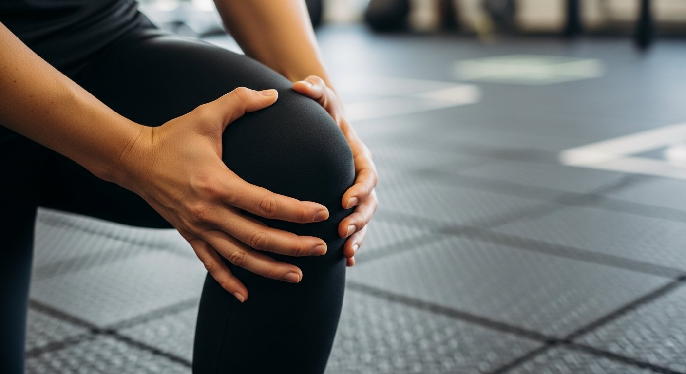
Recognising the symptoms of patellar tendonitis is the first step toward proper diagnosis and treatment.
Pain Location and Character: The most common symptom is pain at the front of the knee, specifically where the patellar tendon attaches to the kneecap or shinbone. This pain typically starts as a dull ache that worsens with activity and improves with rest.
Symptom Progression: Early in the condition, pain may only occur at the beginning of physical activity, subsiding during warm-up, and returning after activity ends. As the condition progresses, pain may persist throughout activity and eventually affect daily movements like climbing stairs or sitting with bent knees.
Physical Signs: The area may become tender to touch, and in some cases, swelling or redness appears around the patellar tendon. Some patients report stiffness, particularly after periods of inactivity, such as waking in the morning.
Functional Impact: Advanced cases may cause weakness in the affected leg, difficulty fully straightening the knee, or a feeling of instability during weight-bearing activities. Without treatment, these symptoms typically worsen over time.
Causes and Risk Factors
The development of patellar tendonitis stems from various factors related to physical activity, biomechanics, and individual characteristics.
Repetitive Stress: The primary cause is repeated strain on the patellar tendon, particularly from activities involving jumping and landing. The condition earned its nickname “jumper’s knee” due to its prevalence in sports like basketball and volleyball.
Biomechanical Factors: Flat feet, knock knees, leg length discrepancies, and muscle imbalances can alter how force travels through the knee, increasing stress on the patellar tendon. Tight quadriceps and hamstring muscles further contribute to this problem.
Training Errors: Sudden increases in training intensity or frequency, inadequate warm-up routines, and training on hard surfaces place excessive strain on the tendon. People who exercise intensely but irregularly, such as those who are only active on weekends, are particularly at risk.
Other Factors: Age-related tendon degeneration, obesity, previous knee injuries, and certain medical conditions like rheumatoid arthritis can increase vulnerability to patellar tendonitis.
Diagnosing Patellar Tendonitis
Proper diagnosis involves several steps to rule out other knee conditions and assess the severity of tendon damage.
Clinical Evaluation: Diagnosis begins with a thorough medical history and physical examination. The doctor will likely ask about activity patterns, symptom onset, and pain characteristics. Physical tests often include the single-leg decline squat, which typically reproduces pain in patients with patellar tendonitis.
Imaging Studies: While X-rays cannot visualise soft tissue like tendons, they help rule out other conditions such as fractures or arthritis. Ultrasound and MRI provide detailed images of the tendon, revealing thickening, tears, or inflammatory changes characteristic of tendonitis.
Differential Diagnosis: Several conditions present with similar symptoms, including patellofemoral pain syndrome, Osgood-Schlatter disease, patellar tracking disorders, and meniscal tears. The diagnostic process aims to distinguish between these possibilities.
Treatment Approaches
Treatment typically follows a stepwise approach, beginning with conservative measures and progressing to more invasive options if necessary.
Initial Management (PRICE Protocol): The first line of treatment includes Protection, Rest, Ice, Compression, and Elevation. Modifying or temporarily ceasing aggravating activities allows the tendon to heal, while ice therapy reduces pain and inflammation. Anti-inflammatory medications may provide short-term symptom relief.
Physiotherapy: A structured rehabilitation programme forms the cornerstone of effective treatment. This typically includes eccentric strengthening exercises, which involve lengthening the tendon under load. Progressive loading helps stimulate tendon remodelling and healing. Stretching exercises for the quadriceps, hamstrings, and calf muscles improve flexibility and reduce strain on the patellar tendon.
Supportive Measures: Patellar tendon straps or braces redistribute force away from the tendon, potentially reducing pain during activity. Proper footwear and orthotic inserts address biomechanical issues that contribute to tendon stress.
Advanced Interventions: For cases resistant to conservative treatment, options include extracorporeal shock wave therapy, which delivers pressure waves to promote healing; platelet-rich plasma injections, which introduce growth factors to the damaged area; and in severe cases, surgical procedures to remove damaged tissue or repair tendon tears.
Prevention Strategies
Preventing patellar tendonitis involves addressing both training practices and biomechanical factors.
Training Modifications: Follow the “10% rule” by increasing training intensity, duration, or frequency by no more than 10% per week. Incorporate adequate warm-up and cool-down routines into every exercise session. Cross-train with low-impact activities like swimming or cycling to reduce repetitive stress on the knees.
Strength and Flexibility: Develop a balanced strengthening programme for the legs, particularly the quadriceps, hamstrings, and calf muscles. Regular stretching maintains flexibility and reduces tendon strain. Core strengthening improves overall biomechanics and posture during athletic movements.
Equipment and Environment: Wear properly fitted footwear appropriate for your specific sport or activity. Consider shock-absorbing insoles for additional impact reduction. When possible, train on forgiving surfaces like rubberised tracks rather than concrete or asphalt.
Recovery Practices: Allow adequate recovery time between intense training sessions. Consider incorporating foam rolling and massage to maintain muscle and tendon health. Maintain proper nutrition and hydration to support tissue repair processes.
When to Seek Medical Attention
Seek medical advice if knee pain persists despite two weeks of rest and self-care measures, or if symptoms worsen or interfere with daily activities. Immediate medical attention is necessary for sudden severe pain accompanied by a popping sensation (which may indicate tendon rupture) or if significant swelling, redness, or warmth develops around the knee. Frequent flare-ups also warrant professional assessment, as they may suggest underlying biomechanical problems or training errors requiring specific intervention.
Conclusion
Patellar tendonitis represents a common but manageable condition affecting the knee. With proper diagnosis, appropriate treatment, and preventive measures, most individuals achieve good outcomes and return to their desired activities. The key lies in early intervention, consistent adherence to rehabilitation protocols, and addressing the underlying factors that contribute to tendon stress.
Schedule a consultation today and take the first step toward overcoming your patellar tendonitis.