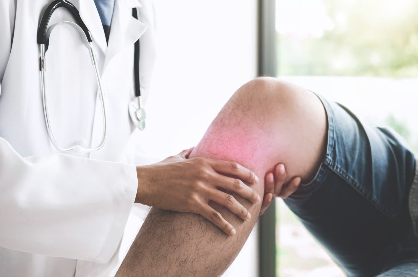
Did you know your knee can grow new bone in response to joint stress? Bone spurs in the knee, medically termed osteophytes, form when the body deposits extra bone along joint edges in response to cartilage breakdown or mechanical stress. These bony projections develop gradually over months to years. They often accompany osteoarthritis (a condition where protective cartilage gradually wears down) as the knee attempts to stabilise itself against deteriorating joint surfaces.
The danger of knee bone spurs depends entirely on their location, size, and interaction with surrounding structures. A small osteophyte tucked along the outer joint margin may never cause problems. However, one growing near the patella or the kneecap, or into the intercondylar notch (the groove between the thigh bone’s lower end) can catch on soft tissues, limit range of motion, or irritate the joint lining.
How Bone Spurs Form in the Knee
Osteophyte formation begins at the cellular level when chondrocytes (cartilage cells) and osteoblasts (bone-forming cells) respond to abnormal mechanical loading. When cartilage thins or sustains damage, stress distribution across the joint changes. Forces concentrate at specific points along bone margins. The body interprets these pressure changes as signals to reinforce the area. This triggers new bone growth.
The process involves several stages. Initial cartilage damage exposes subchondral bone (the layer of bone just beneath the cartilage) to increased stress. Growth factors released during the inflammatory response stimulate mesenchymal stem cells (cells that can develop into different tissue types) in the joint lining and periosteum (the membrane covering bones). These cells differentiate into bone-forming osteoblasts that deposit calcium and phosphate along joint margins. Over time, the soft cartilaginous growth ossifies into mature bone.
Common locations for knee osteophytes include:
- The medial and lateral tibial plateaus (the flat top surfaces of the shin bone)
- The femoral condyles (the rounded ends of the thigh bone)
- The posterior surface of the patella (back of the kneecap)
- The margins of the intercondylar notch
Each location produces distinct mechanical effects and symptom patterns based on proximity to ligaments, tendons, and the joint capsule.
Symptoms That May Indicate Problematic Bone Spurs
Mechanical symptoms occur when osteophytes physically obstruct normal joint movement. Patients describe catching sensations, particularly during the final degrees of knee extension (straightening) or deep flexion (bending). Some experience a hard endpoint when straightening the leg; the knee simply stops short of full extension regardless of effort. Locking episodes, in which the knee temporarily refuses to bend or straighten, suggest an osteophyte catching on soft tissue or an opposing bone.
Pain patterns from bone spurs differ from typical arthritis discomfort. While arthritic pain tends to be diffuse and aching, osteophyte-related pain often localises to specific joint margins. Palpation (gentle pressing) along the joint line may reveal distinct tender spots corresponding to spur locations. Pain that worsens at particular points in the range of motion, rather than throughout movement, suggests mechanical irritation.
Swelling and inflammation develop when osteophytes irritate the synovial lining (the membrane that produces lubricating fluid for the joint). The joint produces excess fluid in response. This creates visible swelling and a sensation of tightness. This reactive synovitis (inflammation of the joint lining) can occur intermittently, flaring after activities that stress the affected area.
Diagnostic Approaches
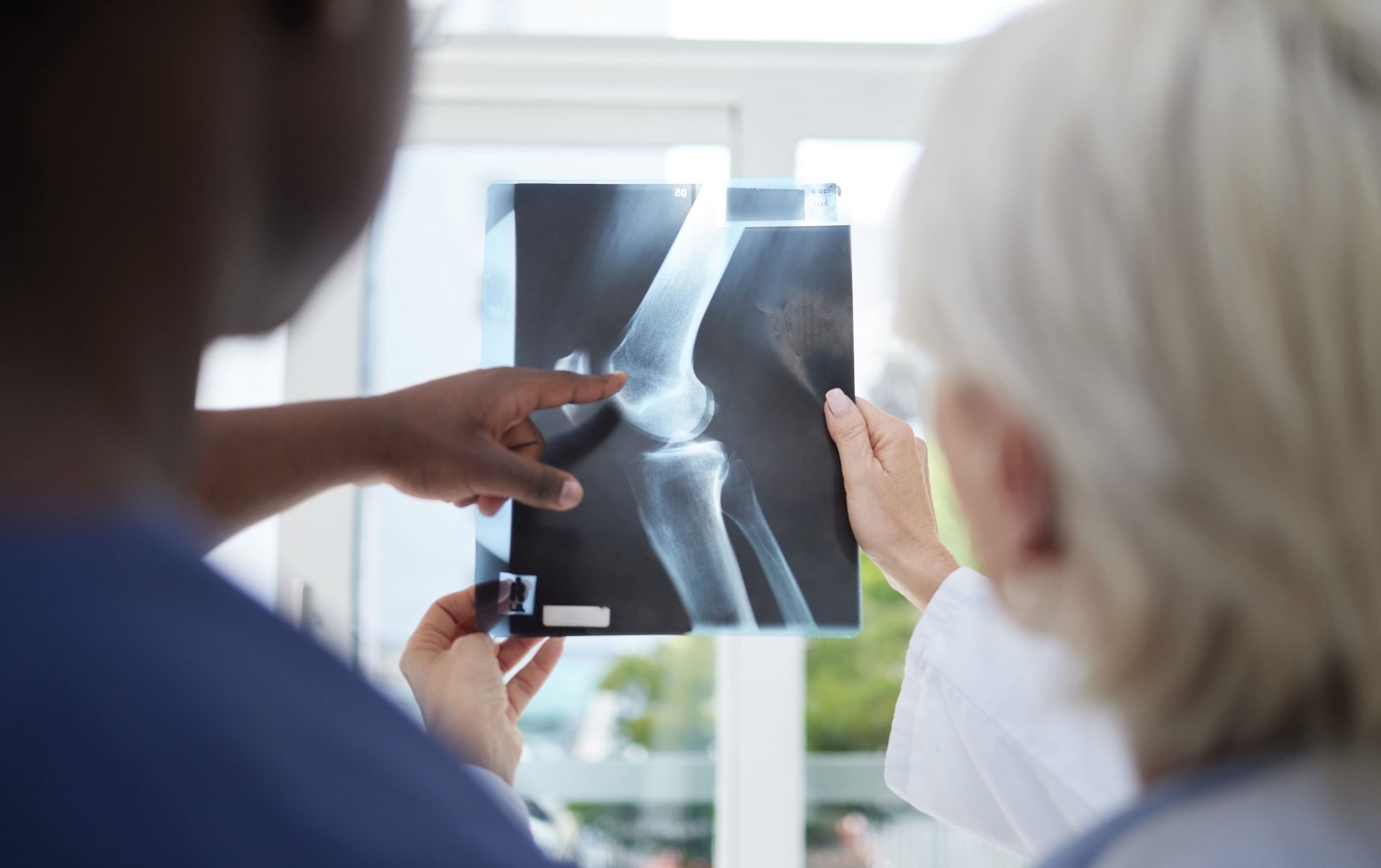
Plain radiographs (X-rays) remain an established screening tool for identifying bone spurs. Weight-bearing X-rays in anteroposterior (front-to-back), lateral (side), and skyline views can show osteophyte location, size, and relationship to the joint space. These images provide valuable information to help your doctor monitor joint health and detect bone changes. Healthcare providers use the Kellgren-Lawrence grading system for osteoarthritis, which incorporates osteophyte presence as a diagnostic criterion.
When symptoms don’t match X-ray findings, additional imaging clarifies the situation. MRI (magnetic resonance imaging, which uses magnetic fields to create detailed pictures of soft tissues and bones) can detect smaller osteophytes that plain films miss. This applies particularly to those developing within the intercondylar notch or along the posterior femoral condyles. MRI also reveals associated soft tissue changes, cartilage damage, meniscal tears (tears in the shock-absorbing cartilage pads), or synovial inflammation that may contribute to symptoms attributed to bone spurs.
CT scanning (computed tomography, which uses X-rays to create cross-sectional images) provides detailed three-dimensional views of osteophyte morphology. Surgeons often request CT imaging when planning arthroscopic removal (a minimally invasive procedure using a small camera and instruments). It precisely maps spur size, shape, and attachment to native bone.
Physical examination findings that suggest symptomatic bone spurs include:
- Loss of terminal knee extension compared to the opposite side
- Palpable hard prominences along joint margins
- Crepitus (grinding or crackling sensation) localised to specific areas rather than generalised grinding
- Reproduction of symptoms at consistent points in the range of motion
When Bone Spurs Require Treatment
The mere presence of osteophytes on imaging does not mandate treatment. Conservative management addresses symptoms without targeting the spurs directly. Activity modification reduces mechanical irritation—avoiding deep squatting, kneeling, or activities that stress the affected joint region. Physical therapy strengthens surrounding muscles to improve joint stability and optimise biomechanics (the way forces act on your body during movement). Anti-inflammatory medications or corticosteroid injections (steroid medicines injected directly into the joint to reduce inflammation) can help reduce synovitis that bone spurs may trigger.
Surgical intervention may be appropriate when conservative measures fail to provide acceptable function and osteophytes demonstrably cause mechanical problems. Arthroscopic osteophyte removal (cheilectomy), a procedure where the surgeon uses small incisions and camera guidance to shave or remove problematic bone growths while preserving healthy tissue, typically involves several weeks of rehabilitation before returning to normal activities.
Specific surgical indications include:
- Fixed loss of extension exceeding a certain degree that limits the function
- Recurrent mechanical symptoms despite adequate conservative treatment
- Osteophytes causing demonstrable impingement (pinching or blocking) on diagnostic imaging
- Preparation for subsequent procedures, such as ligament reconstruction
💡 Did You Know?
Osteophytes actually serve a mechanical purpose; they increase joint surface area and help redistribute forces across damaged cartilage. This explains why bone spurs often accompany rather than precede joint degeneration, acting as the body’s attempt to stabilise deteriorating joints.
Bone Spurs Versus Other Knee Conditions
Several conditions produce symptoms that overlap with osteophyte-related problems. Accurate diagnosis is important for treatment.
Meniscal tears cause catching and locking similar to bone spur impingement. However, meniscal symptoms typically involve a softer, more elastic sensation rather than the hard mechanical block of bone-on-bone contact. Patients with meniscal tears often identify a specific injury that initiated symptoms. Bone spur symptoms develop gradually.
Loose bodies—fragments of cartilage or bone floating within the joint, create unpredictable locking at varying positions. Unlike osteophytes, which catch at consistent points in the range of motion, loose bodies shift position. This causes symptoms that seem to migrate around the knee.
Patellofemoral syndrome produces anterior knee pain (pain at the front of the knee). Patients may attribute this to patellar osteophytes. However, most patellofemoral pain stems from soft tissue imbalances, cartilage irritation, or tracking abnormalities (when the kneecap doesn’t move smoothly in its groove) rather than bone spurs per se.
Synovial plica syndrome involves irritation of embryological remnants of joint tissue (bands of tissue left over from development). Plica bands can catch during movement similarly to osteophytes. However, they are soft tissue structures that don’t appear on X-rays.
Living with Knee Bone Spurs
For those whose bone spurs don’t warrant surgery, long-term management focuses on protecting the joint and maintaining function.
Weight management directly affects mechanical stress across knee surfaces.
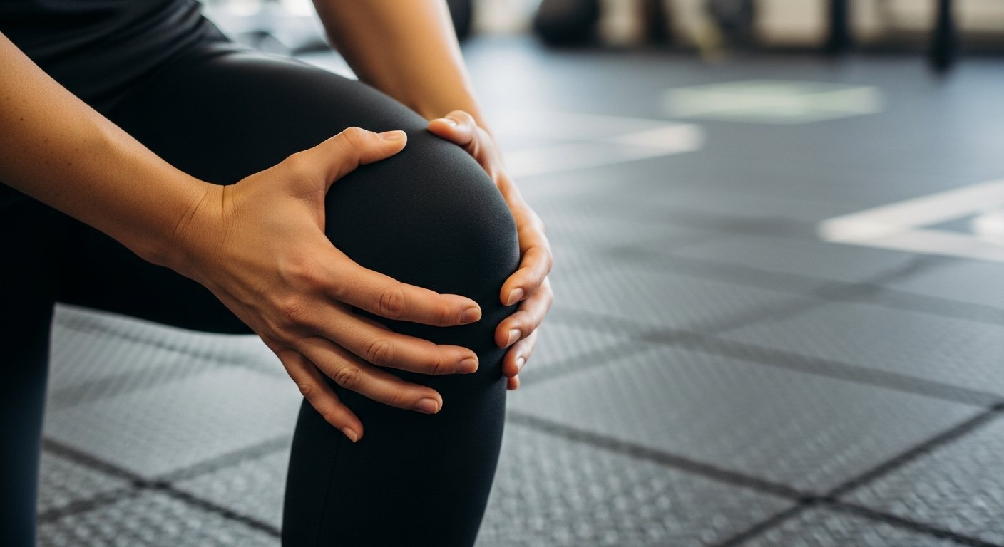
Low-impact exercise helps maintain muscle strength and joint mobility without accelerating degeneration. Swimming, cycling, and elliptical training provide cardiovascular benefits while minimising repetitive impact. Strength training targeting the quadriceps (front thigh muscles), hamstrings (back thigh muscles), and hip muscles supports improved dynamic knee stability.
Activity pacing prevents flares triggered by overuse, rather than alternating between high-activity days and recovery periods. Distributing activities more evenly reduces cumulative joint stress.
Supportive devices may help certain patients. Unloader braces shift weight-bearing forces away from affected compartments. This can potentially reduce osteophyte-related symptoms. Appropriate footwear with adequate cushioning and support protects against ground-reaction forces.
⚠️ Important Note
Bone spurs don’t disappear with conservative treatment; they’re permanent bone changes. However, the inflammation and mechanical irritation they cause often improve with appropriate management, allowing comfortable function despite their radiographic presence.
Progression and Long-Term Outlook
Bone spur growth typically parallels underlying joint degeneration. As cartilage continues to thin and mechanical stresses persist, existing osteophytes may enlarge. New ones may form. However, the response varies considerably between individuals.
Factors associated with slower progression include:
- Maintaining a healthy body weight
- Staying physically active within appropriate limits
- Avoiding high-impact activities that accelerate joint wear
- Addressing biomechanical abnormalities through physical therapy or orthotics (custom shoe inserts or braces that improve alignment)
Factors associated with faster progression include:
- Obesity
- Occupational activities requiring repetitive kneeling or squatting
- Previous knee injuries
- Anatomical malalignment (such as bow-leggedness or knock-knees) that concentrates stress on specific joint areas
Regular clinical monitoring allows early identification of progression. Periodic assessment of range of motion, functional capacity, and symptoms guides decisions about advancing treatment when conservative measures become insufficient.
When to Seek Professional Help
- Pain that interferes with walking, stair climbing, or daily activities despite rest and over-the-counter medications
- Progressive loss of knee straightening compared to your baseline
- Recurrent catching or locking episodes that affect confidence in the knee
- Swelling that persists for more than several days after normal activities
- Night pain that disrupts sleep
- Giving way or instability sensations
- Symptoms that progressively worsen despite activity modification
Commonly Asked Questions
Can bone spurs in the knee go away on their own?
Once formed, bone spurs are permanent structural changes that don’t resolve spontaneously. The bone has fully mineralised into mature tissue. However, symptoms often fluctuate based on activity levels, inflammation, and surrounding tissue health. Periods of minimal symptoms can occur even with unchanged osteophyte size.
Do bone spurs always mean I have arthritis?
Osteophytes most commonly accompany osteoarthritis. However, they can also form after trauma, due to chronic mechanical stress from sports or occupations, or as part of inflammatory conditions. Isolated bone spurs without cartilage damage occur, though this pattern is less common than the combined osteophyte-arthritis presentation.
Will knee replacement remove my bone spurs?
Total knee replacement removes all articular surfaces (joint surfaces), including osteophytes. Surgeons replace them with metal and polyethene (a type of durable plastic) components. Partial knee replacement addresses one compartment while leaving others intact; bone spurs in non-replaced areas would remain.
Can I exercise with bone spurs in my knee?
Exercise remains important for joint health even with osteophytes present. The appropriate activities should maintain strength and mobility without exacerbating symptoms. Low-impact options typically work well. Activities involving deep flexion, pivoting, or impact may require modification. A healthcare professional can help determine suitable exercise options.
How do I know if my symptoms are from bone spurs or something else?
Accurate diagnosis requires clinical examination and imaging correlation. Symptoms occurring at consistent points in range of motion, hard mechanical endpoints, and palpable joint margin tenderness suggest osteophyte involvement. However, multiple conditions often coexist in degenerating knees.

Experiencing Knee Pain or Injury?
Get a Personalised Treatment Plan
Find relief with our hip & knee specialists.
Make An EnquiryConclusion
Bone spurs represent the body’s response to joint stress. Conservative management, including weight control, appropriate exercise, and anti-inflammatory treatments, addresses symptoms in most cases. Surgical removal applies to persistent mechanical problems that limit function despite conservative care.
If you’re experiencing persistent knee pain, catching sensations, or progressive loss of motion, consult an orthopaedic specialist to evaluate your condition and discuss appropriate treatment options.
⚠️ General Disclaimer
Individual recovery experiences, symptom patterns, and treatment responses will vary based on personal health factors, the specific characteristics of the bone spur, and overall joint health. The information provided here is for educational purposes. Consult qualified healthcare professionals for personalised evaluation and treatment recommendations tailored to your specific situation rather than relying solely on general content.