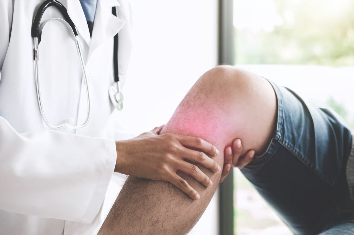
Hamstring injuries near the knee involve the tendons that anchor to the tibia and fibula, creating unique challenges that differ from standard mid-thigh muscle strains. These distal injuries typically manifest as pain behind the knee, difficulty bending the leg, and instability during movements like pivoting or slowing down.
The specific location of the injury dictates the functional impact; lateral-sided damage compromises your ability to push off or control outward rotation, while medial-sided injuries weaken internal stability and the knee’s resistance to inward collapsing forces. Understanding these distinct patterns is essential for accurately diagnosing symptoms and setting realistic expectations for your recovery journey.
Anatomy of the Distal Hamstring Complex
The biceps femoris tendon is the main structure on the outer back of the knee. It attaches mostly to the fibular head and works with the lateral collateral ligament to provide rotational stability.
On the inner side, the semitendinosus joins two other tendons to form the pes anserinus. This broad attachment sits on the tibia a few centimetres below the joint. Nearby, the semimembranosus has a more complex set of attachments that support the knee capsule.
Beyond moving the leg, these tendons provide dynamic stability. They decelerate the leg during your stride and control how the shin bone rotates. If these functions are disrupted, the body adopts compensatory movements that can strain other parts of the knee.
Common Causes and Injury Mechanisms
Acute Avulsion Injuries
Sudden, forceful knee extension against maximum hamstring resistance can avulse tendons from their bony attachments. This mechanism involves the tendon being physically pulled away from the bone where it connects.
Water-skiing injuries are a classic cause of biceps femoris avulsions. This occurs when a ski catches and forces the knee straight while the hamstrings are contracting at their peak. You may see similar injury patterns in football tackles or during heavy gymnastic landings.
Chronic Tendinopathy
Repetitive loading without adequate recovery leads to progressive tendon degeneration rather than sudden failure. Runners and cyclists frequently develop distal hamstring tendinopathy, particularly affecting the biceps femoris insertion. Training errors commonly precede symptom onset. These include:
- Rapid increases in hill work
- Increased speed sessions
- Overall volume increases
Iatrogenic Injury
Anterior cruciate ligament reconstruction using hamstring autografts harvests the semitendinosus tendon and, often, the gracilis tendon. These tendons can regenerate, but the process takes many months. Some patients experience persistent weakness or pain at the harvest site. Improper surgical technique can damage the remaining semimembranosus or cause neurovascular complications.
Direct Trauma
Falls onto the posterolateral knee or direct blows during contact sports can contuse or partially tear the biceps femoris tendon. These injuries often accompany damage to the common peroneal nerve. This nerve wraps around the fibular head immediately adjacent to the tendon insertion.
Recognising Symptoms of Distal Hamstring Injuries
Pain location provides the first diagnostic clue. Biceps femoris pathology causes posterolateral knee pain that may radiate down toward the outer calf. Medial-sided injuries produce pain along the inner knee. This is sometimes confused with pes anserine bursitis or medial meniscus problems.
Swelling patterns differ from typical knee joint effusions. Distal hamstring injuries cause localised swelling directly over the affected tendon rather than generalised knee puffiness. Bruising tracking down the calf suggests more significant structural damage with bleeding into tissue planes.
Functional deficits become apparent during specific activities:
- Difficulty controlling descent on stairs or slopes
- Weakness when pushing off during running or jumping
- Knee giving way during quick direction changes
- Pain with prolonged sitting and the knee bent past a right angle
- Cramping sensation in the posterior thigh during activity
Complete tendon avulsions create palpable gaps or bunching where the retracted muscle belly accumulates. Partial tears produce tenderness directly over the insertion without obvious contour abnormality.
💡 Did You Know?
The semitendinosus tendon can regenerate after surgical harvest for ACL reconstruction. The new tendon forms within the original tendon sheath. However, it typically has a smaller cross-sectional area and different mechanical properties than the original structure.
Diagnostic Approaches
Clinical Examination
Your doctor tests your hamstrings by having you bend your knee against resistance. Testing with your foot turned inward preferentially loads the medial hamstrings. Testing with your foot turned outward isolates the biceps femoris. Comparing strength and pain response between these positions helps localise the injury. Testing in different knee positions helps identify which angle triggers symptoms.
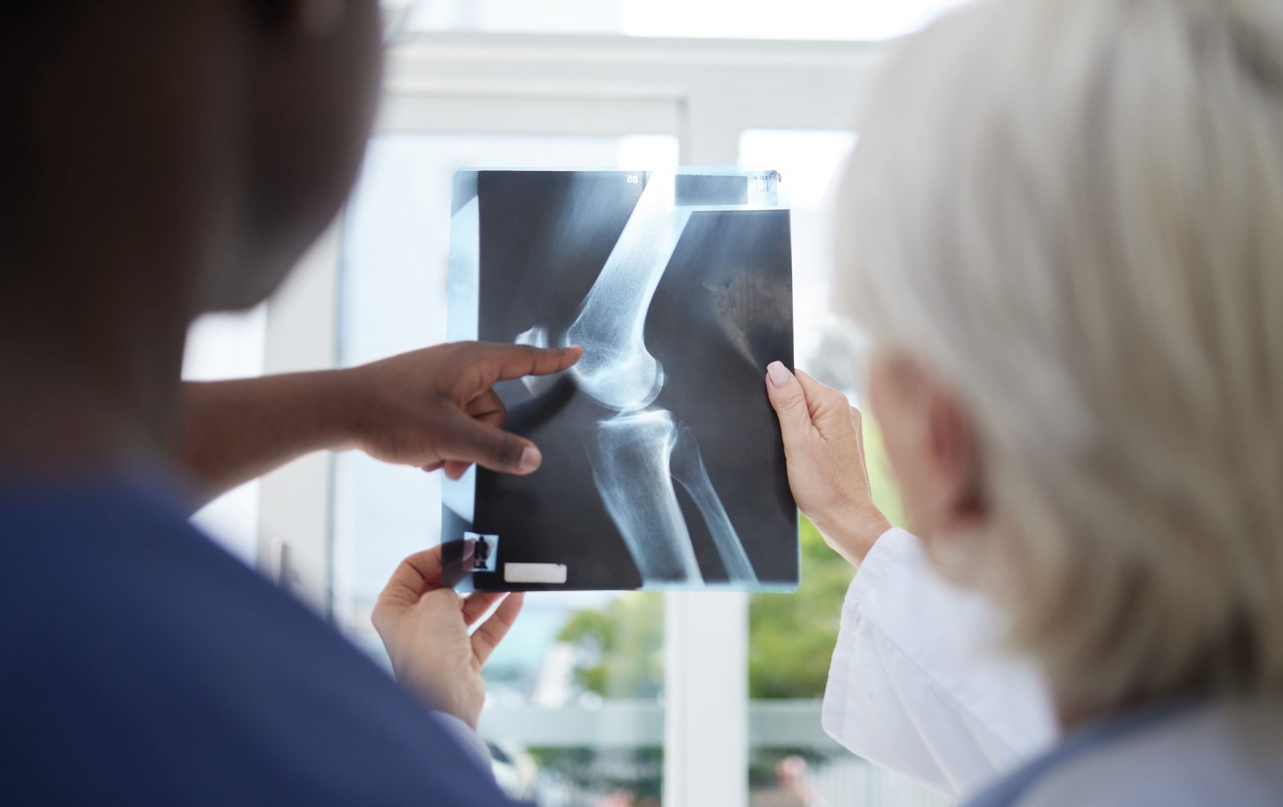
Imaging Studies
MRI provides a detailed look at tendon integrity. It can distinguish between partial and complete tears while measuring how far a tendon has retracted. In chronic cases, the tendon often appears thickened. Acute tears are identified by a clear break in the tissue accompanied by fluid or bleeding.
Ultrasound enables dynamic real-time assessment. This means a clinician can observe the tendons while you move your leg. This method is highly effective for outer structures like the biceps femoris. However, it is less effective for seeing deeper tissues on the inner side of the knee.
Plain radiographs or X-rays are used to find bony avulsions. This occurs when the tendon remains intact but pulls a small piece of bone away from the attachment site. X-rays are essential for confirming injuries to the fibular head or the tibia to determine if surgery is needed.
Treatment Options for Hamstring Tendon Injuries
Non-Operative Management
Many partial tears and tendinopathies respond to structured rehabilitation without surgery. The initial phase focuses on pain control and protecting healing tissue through activity modification rather than complete rest. Early gentle movement prevents adhesion formation and maintains tendon nutrition.
Progressive loading follows specific healing timelines. This process typically begins with isometric exercises within the first two weeks. During this stage, you contract the muscle without moving the joint. The protocol advances to isotonic strengthening by four to six weeks. This phase involves moving the joint through a full range of motion against resistance.
By eight to twelve weeks, you can introduce sport-specific activities. Eccentric exercise programmes may also benefit tendinopathy. In these movements, you focus on controlling the lengthening of the muscle under a load.
Manual therapy techniques, including soft tissue mobilisation and dry needling, may reduce pain and improve tissue extensibility. These interventions work as adjuncts to exercise rather than standalone treatments.
Surgical Intervention
Surgery is often necessary for complete avulsions, especially in active patients or when the tendon has pulled far from the bone. Prompt intervention within three weeks is ideal to prevent scar tissue from making the repair more difficult.
Emergency Evaluation: Biceps femoris tears with nerve symptoms like foot drop require immediate attention. The nearby common peroneal nerve can be compressed by swelling or bleeding.
Fixation Methods: Surgeons use suture anchors or screws to reattach bone fragments in avulsion cases. Mid-substance tears may require a tendon graft if the ends cannot reach each other without too much tension.
Complex Reconstruction: Injuries to the posterolateral corner may use donor tissue to restore stability to the knee.
Debridement: For chronic cases, a surgeon may remove damaged tissue to trigger a fresh healing response. This is sometimes paired with biological treatments like platelet-rich plasma.
Rehabilitation Principles and Timeline
Weeks One to Four: Protection Phase
Weight-bearing progresses as comfort allows. Crutches are used initially for complete tears or surgical repairs. Range of motion exercises maintain joint mobility without stressing healing tissue. Gentle knee bending with gravity assistance avoids active hamstring contraction while preserving movement.
Weeks Four to Eight: Early Strengthening
Isometric exercises (where you contract the muscle without moving the joint) at various knee angles build strength without tendon excursion. Aquatic therapy allows earlier functional movement with reduced loading. Stationary cycling begins once a comfortable range of pain-free flexion is achieved.
Weeks Eight to Twelve: Progressive Loading
Resistance increases systematically. Monitor for delayed pain response that may indicate excessive loading. Single-leg exercises address any asymmetry. Sport-specific movement patterns are introduced at controlled intensities.
Weeks Twelve and Beyond: Return to Activity
Your doctor will set return-to-activity criteria based on your specific injury, recovery progress, and activity goals. Strength testing should show minimal deficit compared to the uninjured side. Functional testing, including hop tests and agility drills, must be completed without pain or apprehension before full activity clearance.
✅ Quick Tip
Nordic hamstring exercises, where you lower yourself forward from a kneeling position while a partner holds your ankles, strengthen hamstrings eccentrically and may help reduce future injury risk once initial healing is complete.
Factors Affecting Recovery Outcomes
Several key variables determine how quickly and successfully you will return to your peak activity level. Understanding these factors helps set realistic expectations for your rehabilitation journey.
Age and Goals: While younger patients often heal faster, older individuals may achieve high satisfaction by meeting lower functional demands.
Tissue Quality: Acute tears in healthy tissue generally repair more reliably than ruptures occurring in already degenerative or thickened tendons.
Protocol Adherence: Following your physical therapy schedule is the strongest predictor of success. Returning to sports too early often leads to re-injury.
Injury Complexity: Recovery is more straightforward for isolated tendon injuries. Combined damage to ligaments, meniscus, or cartilage requires a more complex, integrated treatment plan.
Preventing Hamstring Tendon Injuries
Proactive prevention focuses on preparing the tissue for stress and optimising body mechanics. These strategies help reduce the risk of both sudden tears and chronic overuse injuries.
Dynamic Warm-ups: Use progressive intensity and active stretching to prepare tendons for heavy loads.
Load Management: Monitor weekly training volume and include mandatory recovery days to allow for tissue repair.
Proximal Strengthening: Build hip and core stability to prevent the hamstrings from overcompensating for weak gluteal muscles.
Gait Correction: Use professional movement analysis to identify patterns like overstriding that place excessive stress on the posterior chain.
When to Seek Professional Help
- Posterior knee pain persisting beyond two weeks despite rest
- Sudden pop or tearing sensation followed by weakness
- Visible swelling or bruising behind the knee
- Inability to bend the knee against resistance
- Foot drop or numbness along the outer lower leg (symptoms suggesting nerve involvement)
- Clicking or catching sensations with knee movement
- Progressive worsening despite self-management
- Pain that interrupts sleep or daily activities
Commonly Asked Questions
How long does a distal hamstring injury take to heal?
Recovery timelines vary based on severity and treatment approach, with the degree of improvement varying from person to person. Mild tendinopathy may resolve within six to eight weeks with appropriate rehabilitation. Complete avulsions requiring surgical repair typically need several months before returning to demanding activities. Full tissue maturation continues for up to one year.
Can I continue exercising with a hamstring tendon injury?
Activity modification rather than complete rest usually works. Avoid movements that reproduce pain while maintaining fitness through alternative exercises. Swimming, upper body training, and low-resistance cycling often remain tolerable. Your rehabilitation programme should include progressive loading of the injured tendon once initial inflammation settles.
What’s the difference between hamstring tendinopathy and a tear?
Tendinopathy involves degenerative changes within the tendon substance without structural discontinuity—the fibres remain intact but become disorganised and painful. Tears describe the actual disruption of tendon fibres. These range from minor partial tears to complete ruptures. Imaging studies (such as MRI or ultrasound) differentiate these conditions. Treatment intensity scales accordingly.
Do hamstring tendon injuries require surgery?
Many injuries respond to conservative treatment. Surgery becomes necessary for complete avulsions, particularly with retraction, bony fragments requiring fixation, or associated posterolateral corner instability. Failed conservative treatment after several months of appropriate rehabilitation may also warrant surgical consideration.
Why does my hamstring injury keep coming back?
Recurrence usually indicates incomplete rehabilitation, premature return to activity, or unaddressed contributing factors. Inadequate strength restoration leaves tendons vulnerable to re-injury. Biomechanical issues, training errors, or neural tension problems may perpetuate symptoms. Comprehensive assessment beyond the tendon itself often identifies remediable factors.
Next Steps
Distal hamstring injuries require accurate diagnosis to distinguish tendinopathy from tears and identify associated structures involved. Structured rehabilitation progressively restores function while respecting tissue healing timelines. Your healthcare provider will establish specific treatment goals based on your injury pattern and activity level.
If you’re experiencing posterior knee pain, weakness with bending movements, or instability during physical activities, consult an orthopaedic knee surgeon for a comprehensive evaluation and treatment options tailored to your specific injury.

Experiencing Knee Pain or Injury?
Get a Personalised Treatment Plan
Find relief with our MOH-accredited hip & knee specialists.
Make An Enquiry