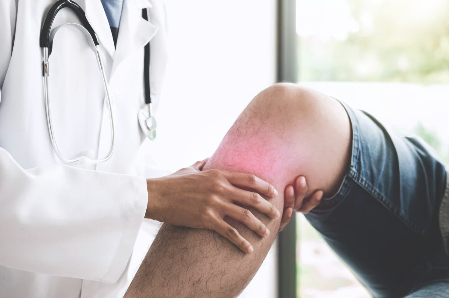
Did you know that the bulge behind your knee might be your body’s warning signal about a deeper joint problem? A Baker’s cyst develops when excess synovial fluid accumulates in the popliteal bursa, which is a small fluid-filled sac located behind the knee. This swelling produces a visible bulge that causes tightness and discomfort during movement, representing a secondary response to an underlying knee joint issue. The bursa communicates with the joint through a one-way valve that allows fluid to enter but prevents it from reentering the joint.
Repeated cycles of fluid production gradually expand the bursa, which explains why these cysts frequently accompany conditions such as osteoarthritis, meniscal tears, and rheumatoid arthritis, which trigger increased fluid production.
How Baker’s Cysts Form
Baker’s cysts form when the delicate balance of fluid production in the knee is disrupted by joint damage or inflammation.
-
Synovial Overproduction: Damage to the joint lining triggers the knee to produce excess lubricating fluid for cartilage nutrition, leading to an accumulation that exceeds the body’s absorption capacity.
-
One-Way Valve Mechanism: A thin channel allows this excess fluid to enter the bursa behind the knee during bending but closes when the leg straightens, effectively trapping the fluid in the sac.
-
Structural Expansion: Continuous accumulation stretches the bursa wall and can lead to internal dividing walls or pockets, occasionally extending into the calf and mimicking blood clot symptoms.
Common Underlying Causes
Osteoarthritis
Degenerative joint changes (gradual wear of joint surfaces) lead to chronic low-grade inflammation in the knee. Cartilage breakdown releases inflammatory mediators (chemical signals) that stimulate synovial fluid production. The worn joint surfaces also directly irritate the synovium (joint lining). Osteoarthritis is a common cause of Baker’s cysts in adults over 50.
Meniscal Tears
Damaged meniscal tissue refers to torn knee cartilage that triggers a reactive joint effusion, a significant fluid buildup in the knee. The tear itself may allow synovial fluid to leak into surrounding structures. Posterior horn tears represent damage to the back part of the meniscus, and those extending to the joint capsule correlate strongly with cyst development. The meniscus normally helps distribute load across the joint, but when it is torn, abnormal stress patterns perpetuate inflammation.
Inflammatory Arthritis
Rheumatoid arthritis is an autoimmune disease that attacks joints, while psoriatic arthritis involves joint inflammation associated with psoriasis, and gout involves crystal deposits that cause joint inflammation. These conditions cause synovial membrane hyperplasia, which is a thickening of the joint lining. The thickened and inflamed synovium produces excess fluid as part of the inflammatory cascade. Inflammatory arthritis can produce particularly large cysts due to the persistent nature of synovial inflammation.
Ligament Injuries
Anterior cruciate ligament tears involve damage to a major knee stabilising ligament, and this, along with other ligament damage, creates joint instability. The resulting abnormal joint mechanics cause repetitive microtrauma, which are tiny repeated injuries to the cartilage and synovium. Post-injury effusion refers to fluid accumulation after injury that often persists for months, providing ongoing fluid for cyst expansion.
Recognising Baker’s Cyst Symptoms
A Baker’s cyst is primarily identified by a soft, fluid-filled swelling in the popliteal fossa, which is the hollow area located directly behind the knee.
-
Mechanical Symptoms: Patients often report tightness or fullness that intensifies when the knee is fully straightened or during prolonged standing, sometimes creating a mechanical block that limits full knee bending.
-
Pain Patterns: While the cyst itself is often painless, underlying joint issues cause aching behind the knee that worsens with activity, occasionally sending referred pain down the calf if the cyst compresses nearby structures.
-
Rupture Risks: If the cyst breaks open, it triggers a sudden onset of severe calf pain, swelling, and bruising as fluid accumulates between muscles, a presentation that closely mimics deep vein thrombosis, which is a dangerous blood clot in the leg.
💡 Did You Know?
Baker’s cysts are named after British surgeon William Morrant Baker, who described them in 1877. He noted the connection between knee joint disease and these popliteal swellings, though the anatomical bursa had been identified decades earlier.
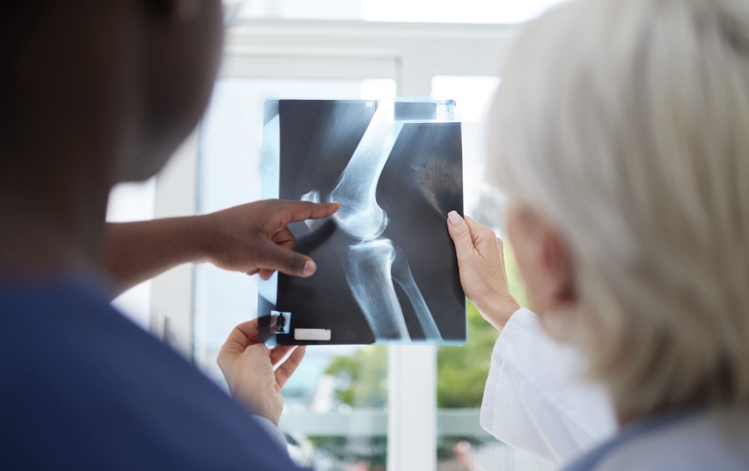
Diagnostic Evaluation
Diagnostic evaluation identifies both the cyst and the underlying joint issue through physical assessment and advanced imaging.
-
Clinical Examination: Physical assessment reveals a mass that firms when the knee is straightened and softens when bent. Transillumination confirms it is fluid-filled by shining a light through the swelling.
-
Ultrasound and Doppler: This ultrasound scan visualises the cyst and rules out a popliteal artery aneurysm, a bulge in the artery behind the knee. A specialised Doppler technique is used to exclude a deep vein thrombosis.
-
Magnetic Resonance Imaging: This detailed MRI scan evaluates the cyst and the underlying joint pathology. It identifies meniscal tears or ligament injuries and helps distinguish simple cysts from complex lesions.
-
Guided Aspiration: A needle is used to withdraw fluid under ultrasound guidance. Analysis of the fluid can reveal inflammatory cells indicating arthritis, blood suggesting injury, or crystals that indicate gout.
Conservative Treatment Approaches
Activity Modification and Rest
Reducing activities that stress the knee decreases fluid production. Avoiding deep squats, prolonged standing, and repetitive impact helps reduce inflammation. Relative rest—maintaining gentle movement while limiting aggravating activities—prevents joint stiffness while promoting healing.
Ice Application
Cold therapy applied to the posterior knee reduces swelling and provides pain relief. Ice packs wrapped in cloth prevent skin damage. Sessions of fifteen to twenty minutes several times daily prove helpful during acute symptom flares.
Compression
Elastic bandages or compression sleeves provide external support and may limit cyst expansion. Compression should be firm but not constricting—avoid the tourniquet effect that impairs circulation. Properly fitted knee sleeves offer convenience for daily wear.
Elevation
Raising the leg above heart level encourages fluid drainage from the knee. Elevation works well when combined with rest periods. Propping the leg on pillows while seated or lying reduces hydrostatic pressure in the joint.
Anti-inflammatory Medications
Nonsteroidal anti-inflammatory drugs (NSAIDs, such as ibuprofen) reduce synovial inflammation and fluid production. Oral medications such as ibuprofen or naproxen provide systemic effects. Topical preparations (creams or gels applied to the skin) offer localised relief with fewer gastrointestinal side effects. The appropriate medication and dosage should be determined by a healthcare professional.
Physical Therapy
Targeted exercises strengthen the muscles that support the knee joint. Stronger quadriceps (front thigh muscles) and hamstrings (back thigh muscles) improve joint stability, reducing abnormal stresses that perpetuate inflammation. Range-of-motion exercises maintain joint flexibility and prevent capsular contracture (stiffening of the joint capsule).
Therapists may employ manual techniques (hands-on treatments) to improve joint mechanics. Patellar mobilisation (gentle movement of the kneecap), soft tissue work, and stretching address contributing factors. A structured programme progresses from gentle movement to functional strengthening over several weeks.
Injection Therapy
Aspiration removes accumulated fluid, providing immediate symptom relief. Ultrasound guidance ensures accurate needle placement within the cyst cavity. Large cysts may require multiple needle positions to achieve complete drainage.
Corticosteroid injection (an anti-inflammatory medication) following aspiration reduces inflammation and slows fluid reaccumulation. The steroid acts on synovial tissue, decreasing its inflammatory activity. Injection into both the cyst and the main knee joint addresses the primary source.
Response to aspiration and injection varies. Some patients experience lasting relief, while others see rapid cyst recurrence. Success correlates with underlying cause—patients with mild osteoarthritis may respond differently than those with significant structural damage or inflammatory arthritis.
Repeated injections carry risks including tendon weakening, skin atrophy (thinning), and potential joint infection. Many clinicians limit corticosteroid injections to a few per year in the same location. A healthcare professional can set treatment targets based on specific risk factors and the underlying knee condition.
⚠️ Important Note
Aspiration without addressing the underlying knee problem typically results in cyst recurrence within weeks to months. The one-way valve mechanism continues pushing fluid into the bursa as long as the joint produces excess fluid.
Surgical Treatment Options
Arthroscopic Treatment
Knee arthroscopy, a procedure in which a surgeon uses a small camera inserted through small incisions to visualise and treat the inside of the joint, addresses internal joint pathology that drives fluid production. The surgeon repairs or trims damaged meniscal tissue, removes loose bodies, and debrides (removes) inflamed synovium. Some techniques include enlarging the valve mechanism to permit bidirectional fluid flow and preventing future accumulation.
Arthroscopy offers minimally invasive access through small incisions. Recovery proceeds faster than open surgery. Many patients return to normal activities within several weeks. Results depend on the adequate treatment of all contributing joint pathologies.
Open Cyst Excision
Direct surgical removal of the cyst, where the surgeon makes an incision to access and remove the entire cyst, may be necessary for large, multiloculated, or recurrent cysts. The procedure requires careful dissection to avoid injury to the popliteal vessels and nerves. Complete excision includes the valve mechanism connecting the cyst to the joint.
Open excision alone, without treating underlying joint pathology, is associated with high recurrence rates. The procedure typically accompanies or follows arthroscopic joint treatment. Post-operative rehabilitation focuses on regaining range of motion and strength.
Combined Approaches
Many surgeons favour combined arthroscopic joint treatment with percutaneous (through the skin) or open cyst decompression. This addresses both the source of fluid production and the accumulation of cysts. Internal valve enlargement during arthroscopy may eliminate the need for direct cyst excision.
What Our Orthopaedic Surgeon Says
Baker’s cysts often frustrate patients because they focus on the visible swelling rather than the underlying knee problem causing it. I explain that the cyst is like the overflow tank for an overproducing joint—draining it helps temporarily, but we need to address why the joint is overproducing fluid. When we successfully treat the meniscal tear, arthritis, or other primary issue, the cyst frequently resolves on its own without direct intervention.
Managing Recovery and Prevention
- Maintain appropriate body weight to reduce mechanical stress on knee joints.
- Continue prescribed exercises after initial treatment. Muscle strength protects joints from excessive stress and improves function regardless of the underlying joint condition.
- Use appropriate footwear with adequate cushioning and support. Worn shoes transmit more impact to the knee joints with each step.
- Address new knee symptoms promptly before chronic effusion develops. Early treatment of meniscal tears and inflammatory arthritis prevents the development of secondary cysts.
- Follow the activity guidelines provided by your treating clinician. Gradual return to activities helps prevent reaggravation of the underlying joint problem.
When to Seek Professional Help
- Sudden severe calf pain with swelling, particularly if the cyst was previously present
- Inability to bear weight on the affected leg
- Numbness or tingling in the foot or lower leg
- Visible skin changes, redness, or warmth over the calf
- Cyst enlarging rapidly over days rather than weeks
- Knee giving way or locking during movement
- Pain interfering with sleep or daily activities despite conservative measures
- Fever accompanying knee or calf symptoms
Commonly Asked Questions
Can a Baker’s cyst resolve spontaneously?
Small cysts may resolve if the underlying knee problem settles spontaneously. However, cysts associated with structural damage, such as meniscal tears or severe osteoarthritis, rarely resolve without treating the underlying condition. Monitoring over several months helps determine whether intervention is necessary.
Is it safe to exercise with a Baker’s cyst?
Low-impact activities like swimming, cycling, and walking typically remain appropriate. High-impact sports and deep squatting may worsen symptoms. A healthcare professional can advise which activities to modify based on specific situations and underlying joint conditions.
How do I know if my Baker’s cyst has ruptured?
Rupture causes sudden, severe pain in the calf with rapid swelling. Bruising may appear around the ankle within a day or two. These symptoms require urgent medical evaluation because they closely resemble deep vein thrombosis, which needs different treatment.
Will the cyst come back after treatment?
Recurrence depends on treatment approach and underlying cause. Simple aspiration without addressing joint pathology is associated with high recurrence rates. Comprehensive treatment, including repair of meniscal tears or management of arthritis, significantly reduces the risk of recurrence.
Can I drain the cyst myself?
Home aspiration is dangerous and not recommended. The cyst lies near major blood vessels and nerves, requiring precise needle placement. Non-sterile technique risks serious infection. Professional aspiration under ultrasound guidance ensures safety.
Next Steps
Baker’s cysts indicate underlying knee joint pathology requiring evaluation. Successful management focuses on identifying and treating the primary condition—whether meniscal damage, arthritis, or other cause. Conservative measures provide relief for many patients, whereas surgical options are available for persistent or complex cases.
If you’re experiencing posterior knee swelling, tightness, or calf pain, consult an orthopaedic surgeon for proper diagnosis and treatment of both the cyst and underlying joint condition.

Experiencing Knee Pain or Injury?
Get a Personalised Treatment Plan
Find relief with our hip & knee specialists.
Make An Enquiry