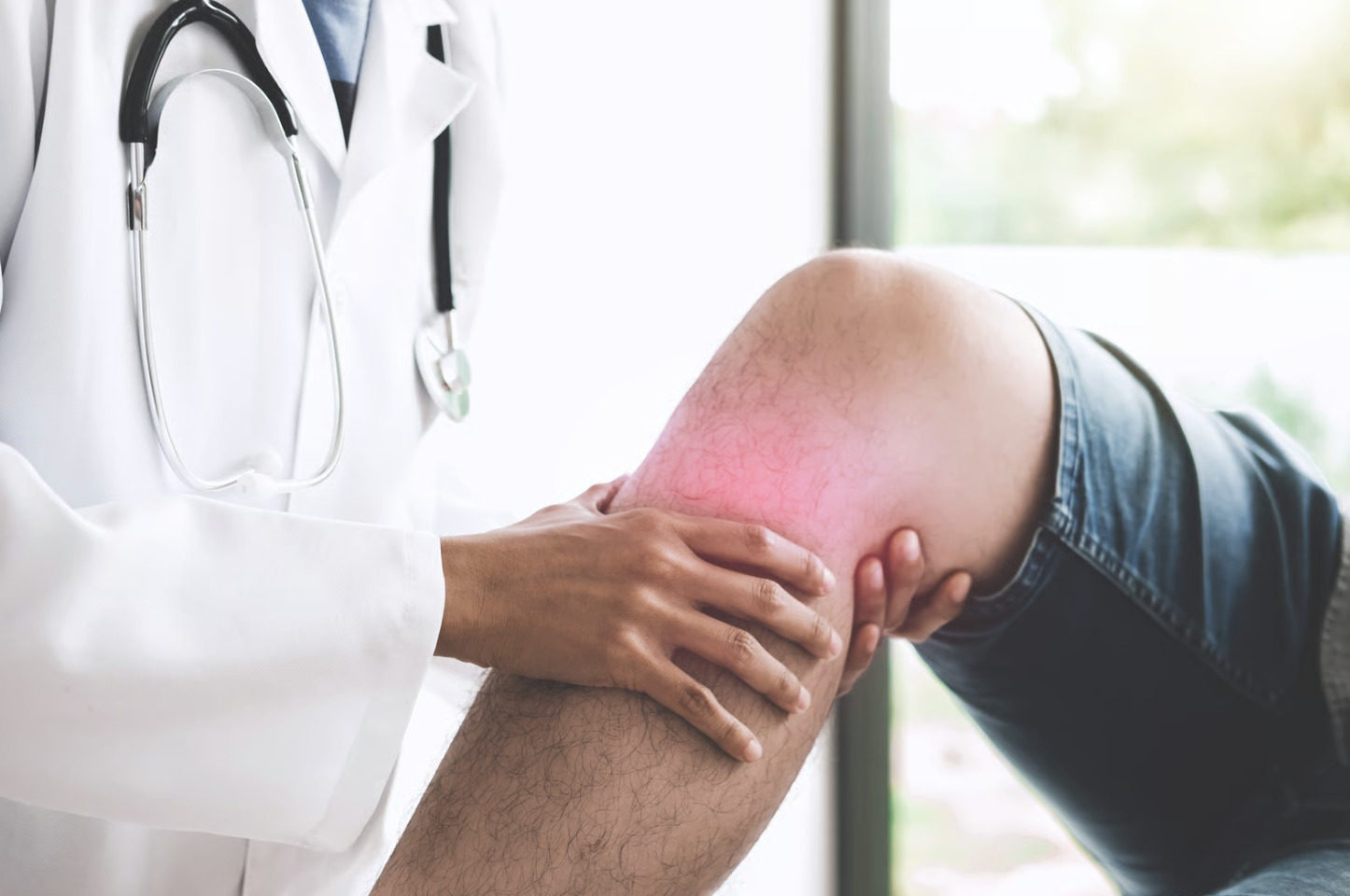
Did you know that your knee joint produces excess synovial fluid when inflamed, leading to the visible swelling that restricts movement? Knee swelling and stiffness signal underlying joint problems that range from acute injuries (such as torn ligaments or meniscus tears) to degenerative conditions (such as osteoarthritis).
The knee joint contains synovial fluid, a lubricating liquid that allows smooth movement. However, inflammation triggers excess fluid production. This causes visible swelling and restricted motion.
Different conditions present distinct patterns:
- Sudden swelling after trauma may suggest injury
- Gradual morning stiffness may indicate arthritis
The location of swelling also provides diagnostic clues:
- Diffuse swelling (swelling throughout the knee) may suggest internal joint problems
- Localised swelling (swelling in one specific area) may point to specific structural damage
Osteoarthritis and Degenerative Changes
Osteoarthritis develops when cartilage (the smooth, protective tissue covering the ends of bones) protecting the knee bones gradually wears away, exposing underlying bone surfaces. This degeneration causes bone-on-bone contact during movement. The contact triggers inflammation and synovial fluid accumulation (a build-up of the lubricating fluid inside the joint). The condition affects older adults, though previous injuries can accelerate development in younger individuals.
Morning stiffness that lasts only a brief period characterises osteoarthritis. It improves with gentle movement as synovial fluid redistributes. Swelling fluctuates with activity levels. Prolonged standing or walking increases fluid accumulation, whilst rest reduces it. The medial (inner) knee compartment commonly degenerates due to increased load bearing during walking.
Bone spurs (osteophytes) form along joint margins as the body attempts to stabilise the damaged joint. These bony projections further restrict movement and contribute to the grinding sensation patients describe. Advanced osteoarthritis shows visible joint deformity. The leg may appear bowed inward or outward, depending on which compartment deteriorates faster.
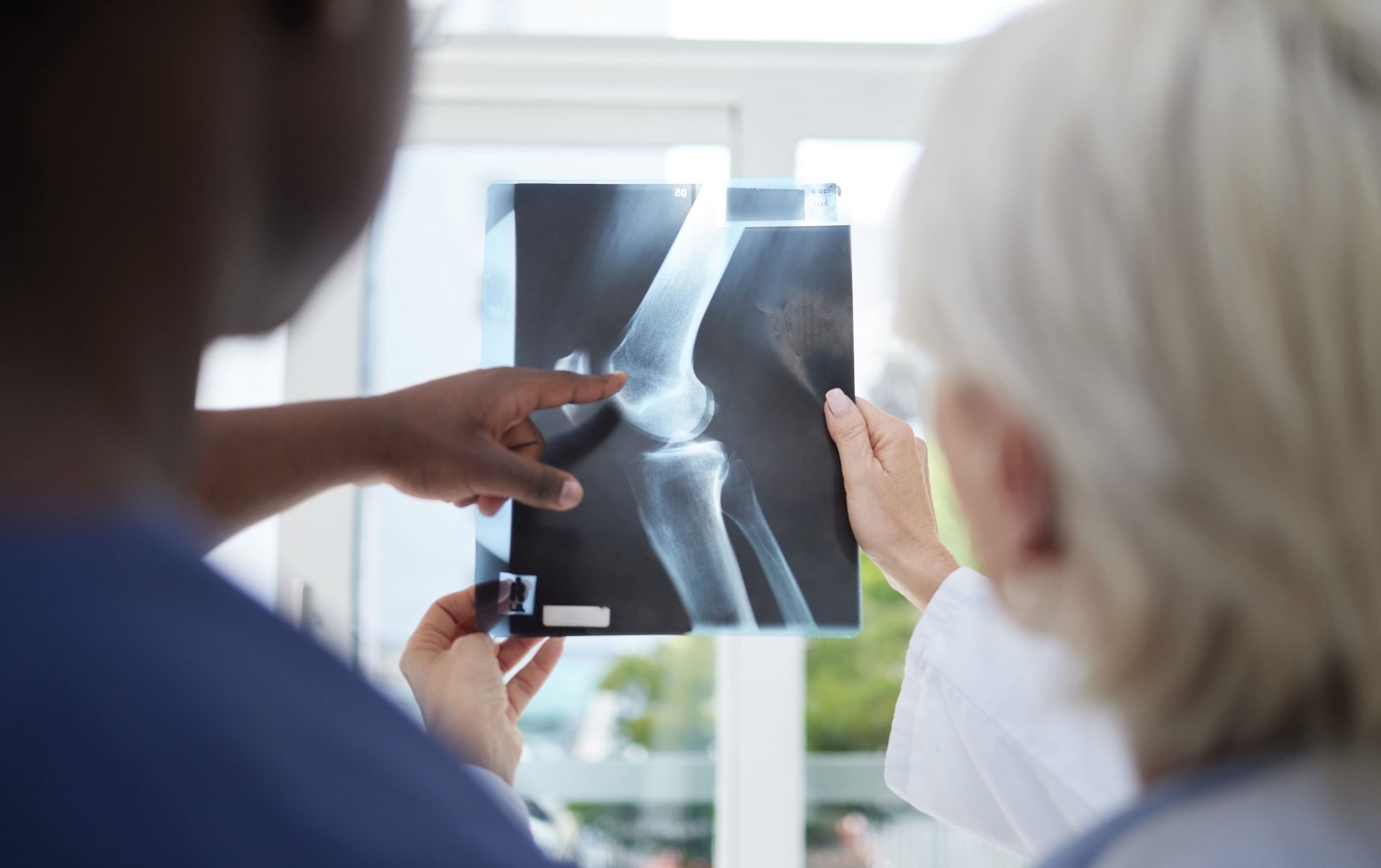
X-rays can reveal several changes:
- Joint space narrowing (the gap between bones becomes smaller as cartilage wears away)
- Subchondral sclerosis (hardening of the bone just beneath the cartilage)
- Osteophyte formation (bone spurs)
MRI can assess cartilage thickness and detect early changes before X-ray abnormalities appear. Weight management can help reduce joint loading.
Meniscus Tears and Cartilage Damage
The meniscus consists of two C-shaped fibrocartilage structures (tough, flexible tissue that acts as a shock absorber) that cushion and stabilise the knee joint. Tears occur through acute twisting injuries or gradual degeneration. Different tear patterns affect symptoms and treatment options:
- Horizontal tears run parallel to the tibial plateau (the flat top surface of the shinbone)
- Radial tears extend from the inner margin outward
Acute meniscus tears produce immediate swelling within a day as blood vessels at the outer meniscal rim bleed into the joint. Patients experience catching or locking sensations when torn fragments interfere with joint movement. The knee may suddenly give way during weight-bearing activities, particularly when rotating.
Degenerative tears develop gradually without specific injury. These tears rarely cause dramatic locking but produce persistent aching and intermittent swelling. Complex tears involving multiple planes create unstable flaps that irritate surrounding tissues with each movement.
Physical examination reveals joint line tenderness along the affected meniscus. The McMurray test can reproduce pain and clicking when the examiner rotates the tibia (shinbone) whilst extending the knee. MRI (a type of imaging scan that creates detailed pictures of internal structures) identifies tear location, pattern, and associated cartilage damage:
- Peripheral tears in the vascular zone (the outer area with blood supply) may heal with conservative treatment
- Central tears in the avascular zone (the inner area without blood supply) typically require surgical intervention
Ligament Injuries and Instability
The anterior cruciate ligament (ACL) prevents forward sliding of the shin bone (tibia). It most commonly tears during sudden direction changes or during landing from jumps. Complete ACL ruptures produce immediate, severe swelling as blood from torn ligament vessels fills the joint cavity within several hours. The knee feels unstable during pivoting movements. Patients describe a sensation of the knee “giving out.”
Medial collateral ligament (MCL) injuries occur from direct blows to the outer knee, causing pain along the inner knee. Grade I sprains involve microscopic tearing with minimal swelling, whilst Grade III tears show complete ligament disruption with significant medial joint opening. MCL injuries often accompany ACL tears in combined injury patterns.
Posterior cruciate ligament (PCL) injuries result from dashboard impacts or falls onto a flexed knee (such as when the bent knee hits the dashboard in a car accident). Swelling develops more gradually than ACL tears, with posterior knee pain predominating. The posterior drawer test (a physical examination where the doctor gently pushes the shin bone backward to check for abnormal movement) demonstrates increased backward movement of the shin bone compared to the uninjured side.
Chronic ligament insufficiency (when the ligament remains weak or damaged over time) leads to recurrent swelling episodes as the unstable joint experiences repetitive microtrauma (minor injuries from everyday movements). Secondary osteoarthritis (joint degeneration and cartilage breakdown) may develop following untreated ACL tears due to altered joint mechanics. Functional bracing can provide external stability during activities. However, it cannot restore standard movement patterns.
Inflammatory Conditions
Rheumatoid arthritis affects the synovial membrane (the thin tissue lining the joint). It causes symmetric joint inflammation that typically involves both knees. Morning stiffness persists for extended periods. Warmth and redness accompany swelling. The inflammatory process erodes cartilage and bone. Without treatment, this can lead to joint deformity.
Gout results from uric acid crystal deposition within the joint. These crystals trigger intense inflammatory responses. Attacks develop suddenly, often overnight. The tenderness becomes so severe that even a light touch is painful. The knee appears red and hot. Swelling develops rapidly over hours. Elevated serum uric acid levels increase the risk of crystallisation, though levels may normalise during acute attacks.
Pseudogout involves calcium pyrophosphate crystal deposition. It affects larger joints in older adults. Symptoms mimic gout but develop more gradually over days rather than hours. X-rays show chondrocalcinosis or linear calcification within cartilage structures. Joint fluid analysis under polarised light microscopy can differentiate crystal types.
Reactive arthritis develops several weeks after gastrointestinal or genitourinary infections. Knee involvement is frequently seen. The condition affects young adults predominantly. Asymmetric joint patterns distinguish it from rheumatoid arthritis. HLA-B27 genetic marker testing can identify predisposition in recurring cases.
Baker’s Cyst Formation
Baker’s cysts develop when excess synovial fluid (the lubricating liquid naturally present in joints) accumulates in the popliteal bursa (a small fluid-filled sac) behind the knee. These fluid-filled sacs form secondary to internal knee problems that increase synovial fluid production. A one-way valve mechanism allows fluid entry but prevents drainage. This causes progressive cyst enlargement.
Cysts produce posterior knee fullness and tightness. This is particularly noticeable when extending the leg. Large cysts restrict knee flexion (bending of the knee) and create visible bulging in the popliteal fossa (the area at the back of the knee). Standing and walking increase discomfort as muscle contraction compresses the cyst.
Cyst rupture releases synovial fluid into the calf. This causes sudden pain, swelling, and bruising that mimics deep vein thrombosis (a blood clot in a deep vein). The “crescent sign” – bruising around the ankle – appears within a few days after rupture as fluid tracks downward. Ultrasound imaging (a scan that uses sound waves to create pictures of the inside of your body) distinguishes cysts from vascular problems and identifies communication with the joint space.
Treatment addresses the underlying joint pathology rather than the cyst itself. Aspiration (a procedure in which the doctor drains fluid from the cyst using a needle) provides temporary relief, but cysts typically recur unless the underlying cause is addressed. Surgical excision (removal of the cyst) combined with closure of the communication channel can help prevent recurrence in persistent cases.
⚠️ Important Note
Baker’s cysts in children often occur without underlying joint pathology and may resolve spontaneously over time, unlike adult cysts, which are usually indicative of internal knee problems requiring evaluation.
Infection and Septic Arthritis
Septic arthritis represents a medical emergency requiring immediate treatment to prevent permanent joint damage. Bacteria enter the joint through:
- Bloodstream spread
- Direct penetration (such as through a wound or injury that reaches the joint)
- Extension from adjacent infections
Staphylococcus aureus (a common skin bacterium) causes many cases. Streptococcal species and gram-negative bacteria occur less frequently.
Infected knees show rapid swelling development over hours to days. This occurs alongside:
- Fever
- Severe pain
- Inability to bear weight
The joint feels hot with overlying skin redness. Any knee movement triggers intense pain. This distinguishes septic arthritis from other inflammatory conditions, in which gentle movement provides relief.
Joint aspiration (a procedure where the doctor uses a needle to withdraw fluid from the joint) reveals cloudy or purulent synovial fluid (the fluid that normally lubricates the joint) with substantially elevated white blood cell counts. Gram staining (a laboratory technique used to identify bacteria) can identify organisms in some cases. Cultures confirm bacterial species and antibiotic sensitivities. Blood cultures may remain positive in patients with septic arthritis.
Delayed treatment can lead to cartilage destruction within days. Bacterial enzymes and inflammatory mediators damage joint structures during this time. Healthcare professionals start empiric antibiotic therapy (treatment that begins before they identify the exact bacteria) immediately after cultures are obtained. They subsequently adjust treatment based on sensitivity results. Serial aspirations (repeated fluid removal procedures) or arthroscopic irrigation (a method where the doctor uses a small camera and instruments to flush out the joint) remove inflammatory debris and monitor treatment response.
Daily Management Strategies
- Apply cold therapy during acute swelling episodes using ice packs wrapped in cloth for short intervals. Cold constricts blood vessels, reducing fluid accumulation and numbing pain receptors. Alternate with gentle movement to prevent stiffness.
- Perform range-of-motion exercises to maintain joint flexibility and promote synovial fluid circulation. Heel slides, seated knee extensions, and gentle stretching can help prevent adhesion formation during healing.
- Modify activities by replacing high-impact exercises with swimming or cycling that maintain fitness without excessive joint stress. Use appropriate footwear with adequate cushioning and arch support to support lower limb alignment.
- Elevate the affected leg above heart level when resting to promote fluid drainage through lymphatic channels. Position pillows under the calf rather than directly under the knee to avoid prolonged flexion.
- Strengthen surrounding muscles through isometric quadriceps contractions and straight leg raises to help improve joint stability without aggravating swelling.
When to Seek Professional Help
- Sudden severe swelling with inability to bear weight
- Fever accompanying knee swelling and pain
- Knee locking in a bent or straight position (the knee gets stuck and won’t move)
- Visible deformity or knee giving way repeatedly
- Swelling persists beyond a considerable period despite rest and ice
- Progressive worsening of symptoms over an extended time
- Night pain is disrupting sleep consistently
- Skin colour changes or warmth that may indicate infection
- Previous knee surgery with new swelling development
Commonly Asked Questions
How long does knee swelling typically last after injury?
Acute injury swelling peaks within the first couple of days. It then gradually resolves over several weeks with appropriate treatment. Persistent swelling beyond this timeframe suggests ongoing inflammation requiring medical evaluation. Complete ligament tears or significant cartilage damage may cause intermittent swelling for months.
Can knee swelling resolve without treatment?
Minor swelling from overuse often resolves with rest, ice, and elevation over several days. However, swelling caused by structural damage, arthritis, or infection requires specific treatment. Untreated mechanical problems can lead to recurring episodes of swelling and accelerated joint degeneration.
What sleeping positions help reduce knee stiffness?
Sleep with a small pillow under the knee to maintain slight flexion. This can help prevent prolonged extension, which can increase morning stiffness. Side sleepers should place a pillow between their knees to maintain alignment. Avoid sleeping with your knees fully bent, as this restricts circulation and increases swelling.
When should compression be avoided for swollen knees?
Avoid compression with suspected infection, deep vein thrombosis, or peripheral vascular disease. Excessive compression impairs circulation and may worsen swelling. Properly fitted compression sleeves provide support without restricting blood flow, with comfortable snugness that doesn’t leave indentations.
Does the weather affect knee swelling and stiffness?
Barometric pressure changes before storms may increase joint discomfort in some arthritis patients. Cold weather increases synovial fluid viscosity, contributing to morning stiffness. While everyone experiences weather sensitivity differently, maintaining consistent indoor temperature and humidity can help minimise symptom fluctuations.
Next Steps

Experiencing Knee Pain or Injury?
Get a Personalised Treatment Plan
Find relief with our hip & knee specialists.
Make An EnquiryAcute injuries with sudden swelling require immediate evaluation to prevent secondary damage. Gradual symptoms often indicate degenerative changes that are manageable through targeted therapy and lifestyle modifications. Early intervention preserves joint function long-term.
If you’re experiencing persistent knee swelling, catching sensations, or joint instability, consult an orthopaedic surgeon to evaluate your condition and discuss treatment options.