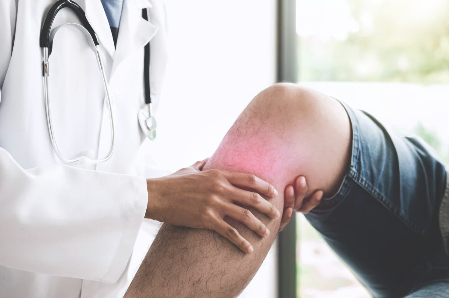
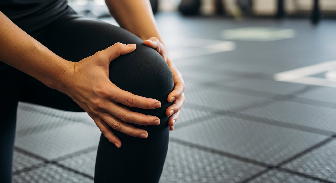
Have you ever felt your knee suddenly freeze mid-movement, unable to bend or straighten? Knee locking occurs when your knee joint becomes stuck in one position, preventing regular bending or straightening. This condition can be divided into distinct types: mechanical locking, in which physical structures (such as torn cartilage or loose bone fragments) block joint movement, and pseudo-locking, in which pain and muscle spasms create the sensation of being stuck. The difference between these types determines both the urgency of treatment and the underlying cause of your knee joint problem.
Mechanical locking typically happens suddenly during movement. It leaves your knee fixed at a specific angle until it is manually released or spontaneously released. Pseudo-locking develops gradually, with increasing pain and stiffness that mimic actual locking but allow some movement when the pain subsides.
Mechanical vs Pseudo-Locking
Mechanical locking results from physical obstruction within the knee joint space. Torn meniscus fragments (pieces of the cushioning cartilage between your thighbone and shinbone), loose cartilage pieces, or bone fragments physically wedge between joint surfaces. This blocks the knee’s normal gliding motion. Your knee stops abruptly mid-movement, often accompanied by a catching sensation. Complete inability to extend or flex the joint follows. The locked position remains fixed regardless of rest or pain medication until the obstruction dislodges.
Mechanical causes include:
- Bucket-handle meniscus tears (where the torn portion flips into the joint space like a bucket handle)
- Osteochondral fragments (pieces of bone and cartilage) from previous injuries are breaking free
- Synovial plica syndrome (where thickened tissue bands catch during movement)
Each mechanical block creates a characteristic pattern: a sudden onset during specific movements, a defined angle at which locking occurs, and immediate relief once the obstruction moves.
Pseudo-locking presents differently through protective muscle guarding rather than physical blockage. Severe arthritis (joint inflammation and damage), patellofemoral pain syndrome (pain around the kneecap), or acute ligament injuries trigger intense pain signals. These signals cause surrounding muscles to contract involuntarily. Your quadriceps and hamstrings simultaneously tighten. This creates functional immobility that feels like actual locking. Unlike mechanical locking, gentle passive movement by a healthcare provider often reveals that the joint can move despite the sensation of being stuck.
The distinction matters for treatment planning. Mechanical locking may require arthroscopic surgery (a procedure where a surgeon inserts a small camera and instruments through tiny incisions) to remove or repair the obstructing tissue. Pseudo-locking responds to pain management, physical therapy, and treating the underlying inflammatory condition and attempting forceful manipulation of a mechanically locked knee risks further damage. Appropriate stretching and strengthening exercises can help resolve pseudo-locking episodes.
Meniscus Tears and Knee Locking
Meniscus tears represent a common cause of actual mechanical knee locking. The meniscus is a C-shaped cartilage cushion between your thighbone and shinbone. It can tear during twisting movements or as a result of degenerative changes. Specific tear patterns create different locking mechanisms:
- Radial tears (straight tears extending from the inner edge outward) rarely lock
- Bucket-handle tears (vertical tears that create a flap of tissue) frequently cause complete mechanical blocks
Bucket-handle tears occur when a vertical tear extends through the meniscus body. This creates a mobile fragment that flips into the joint centre like a bucket handle. The displaced tissue wedges between the femur (thighbone) and tibia (shinbone) during knee bending or straightening. This stops movement completely. The locked position typically occurs at a partially bent angle. It prevents complete straightening. Patients often report hearing a pop at the time of the initial injury, followed by immediate swelling and subsequent locking episodes.
Complex tears combining horizontal and vertical components create unpredictable locking patterns. The torn fragments move within the joint during different activities. This causes intermittent catching that progresses to complete locking. Degenerative tears in older adults develop gradually through repetitive microtrauma (minor repeated injuries). They produce frayed edges that intermittently catch rather than sudden, complete blocks.
Treatment depends on tear location, pattern, and patient factors. Peripheral tears within the vascular zone (the outer area with blood supply) may heal with conservative management. This includes activity modification and structured rehabilitation. Central tears in the avascular white zone (the inner area without blood supply) may require arthroscopic partial meniscectomy or repair. Arthroscopic partial meniscectomy is a procedure where healthcare professionals use a small camera and instruments to remove the damaged portion of the meniscus. Repair involves stitching the tear back together. Locked bucket-handle tears may need arthroscopic reduction. During this procedure, the surgeon repositions the flipped tissue back to its normal location. Post-surgical rehabilitation focuses on restoring the range of motion, strengthening the surrounding muscles, and gradually returning to activities.
Loose Bodies in the Joint
Loose bodies floating within the knee joint create unpredictable locking episodes when they lodge between articulating surfaces (the parts of the joint that move against each other). These fragments originate from various sources:
- Osteochondritis dissecans (a condition where bone and cartilage separate from the underlying tissue)
- Traumatic fractures producing free-floating chips
- Arthritis generates cartilage debris
Each fragment acts as a mechanical wedge that randomly blocks normal joint motion.
Osteochondritis dissecans affects adolescents and young adults. It causes segments of bone and overlying cartilage to lose blood supply and potentially detach. The separated fragment initially remains partially attached, creating catching sensations. It then completely detaches, becoming a loose body. These fragments vary in size. Smaller fragments cause intermittent catching, while larger pieces produce sudden, complete locking.
Synovial chondromatosis (a condition where the joint lining produces abnormal cartilage growths) presents a pattern where the joint lining produces multiple cartilage nodules that break free into the joint space. These nodules may calcify over time, becoming visible on standard X-rays. Patients experience numerous loose bodies, leading to frequent locking episodes at varying knee positions. The condition progresses without treatment, generating increasing numbers of loose bodies that compound mechanical symptoms.
Arthroscopic removal (a minimally invasive surgical procedure using a small camera and instruments) provides treatment for symptomatic loose bodies. The surgeon visualises the entire joint space and removes all accessible fragments while addressing the underlying source when possible. Significant osteochondral defects (damaged areas where both bone and cartilage are affected) may require additional procedures like microfracture (a technique that stimulates new cartilage growth) or cartilage grafting (transplanting healthy cartilage tissue). Post-operative recovery focuses on preventing adhesions (scar tissue that can restrict movement) while protecting healing tissue. This is followed by progressive strengthening to help prevent future fragment formation.
Arthritis-Related Locking
Arthritis creates sensations similar to locking through multiple mechanisms beyond simple pain. Bone spurs (extra bone growth along joint edges) develop along joint margins. They potentially press on surrounding soft tissues during movement. Severe cartilage loss (when the protective cushioning between bones wears away) causes bone-on-bone contact. This triggers protective muscle spasms (involuntary muscle tightening to protect the joint). Inflammatory synovitis (swelling of the joint lining) produces joint effusion (fluid build-up in the joint). This mechanically limits motion while triggering pain-mediated guarding (muscle tightening in response to pain).
Osteoarthritis progression follows predictable patterns affecting locking symptoms. Early-stage disease causes morning stiffness that improves with movement. Moderate arthritis produces catching sensations as roughened cartilage surfaces interact. Severe arthritis can cause pseudo-locking episodes (sensations of the joint being stuck, without an actual mechanical blockage). Inflammation and muscle guarding prevent movement despite the absence of an actual mechanical block. The knee feels stuck in slight flexion, particularly after prolonged sitting or upon waking.
Inflammatory arthritis, including rheumatoid arthritis (where the immune system attacks joint linings) and psoriatic arthritis (arthritis associated with the skin condition psoriasis), creates additional locking mechanisms. Active synovitis thickens the joint capsule (the tissue surrounding the joint). This physically restricts movement. Pannus formation (abnormal tissue growth within the joint) acts similarly to loose bodies when pieces break free. Morning locking episodes lasting for extended periods suggest an inflammatory rather than mechanical cause. These require different treatment approaches.
Management strategies target both mechanical and inflammatory components:
- Intra-articular corticosteroid injections (injections of anti-inflammatory medication directly into the joint) can reduce synovitis and associated pseudo-locking
- Viscosupplementation with hyaluronic acid (injections that add lubricating fluid to the joint) can improve joint lubrication, reducing catching sensations
- Physical therapy helps maintain the range of motion while strengthening muscles to offload arthritic joint surfaces
- Cases with true mechanical blocks from large osteophytes may require surgical debridement (a procedure where the surgeon removes damaged tissue and bone spurs) or total knee replacement (surgery to replace the damaged joint with an artificial one)
Associated Symptoms
Knee locking rarely occurs in isolation. It presents alongside symptoms that help identify the underlying cause.
Swelling patterns provide diagnostic clues. Immediate swelling after injury may suggest haemarthrosis (bleeding into the joint) from ligament or meniscus tears. Gradual swelling over hours can indicate synovial irritation (inflammation of the joint lining). Recurrent effusions (fluid build-up) between locking episodes may point towards ongoing mechanical irritation from loose bodies or unstable meniscus tears.
Pain characteristics differ between mechanical and inflammatory causes. Sharp, localised pain during specific movements may suggest mechanical impingement (when structures get pinched or caught). Diffuse aching that worsens with weather changes can indicate arthritic involvement. Night pain disrupting sleep warrants investigation for inflammatory arthritis or infection. Pain relief with rest supports mechanical causes. Morning stiffness that improves with activity may suggest an inflammatory condition.
Instability sensations accompany locking when ligament insufficiency contributes to abnormal joint mechanics. The knee may buckle or give way during weight-bearing, particularly on uneven surfaces or stairs. ACL tears (injuries to the anterior cruciate ligament, a stabilising structure) allow abnormal anterior translation (forward sliding of the shin bone) that positions the meniscus for impingement. MCL injuries (damage to the medial collateral ligament on the inner knee) create a medial opening that alters load distribution, accelerating meniscus degeneration. Combined injuries result in complex mechanical dysfunction, requiring individualised treatment.
Mechanical symptoms, including clicking, popping, and grinding, provide information:
- Painful clicking with locking may suggest meniscus pathology (damage to the cushioning cartilage)
- Painless popping may indicate normal tendon movement over bone prominences
- Grinding sensations (crepitus) with locking episodes may suggest cartilage loss
Recording when symptoms occur—during specific activities, positions, or times of day—helps clinicians identify patterns pointing to particular diagnoses.
Diagnostic Approaches
Clinical examination combines patient history with specific physical tests to differentiate locking causes. McMurray’s test reproduces meniscus-related clicking and locking by rotating the shin bone whilst bending and straightening the knee. Thessaly’s test, performed whilst standing with knee rotation, often provokes locking in unstable meniscus tears. Joint line tenderness localised to specific areas correlates with the location of a meniscus tear. Effusion assessment using the patellar tap test (a gentle test in which the doctor presses on the kneecap to feel for fluid beneath) quantifies fluid accumulation, suggesting ongoing irritation.
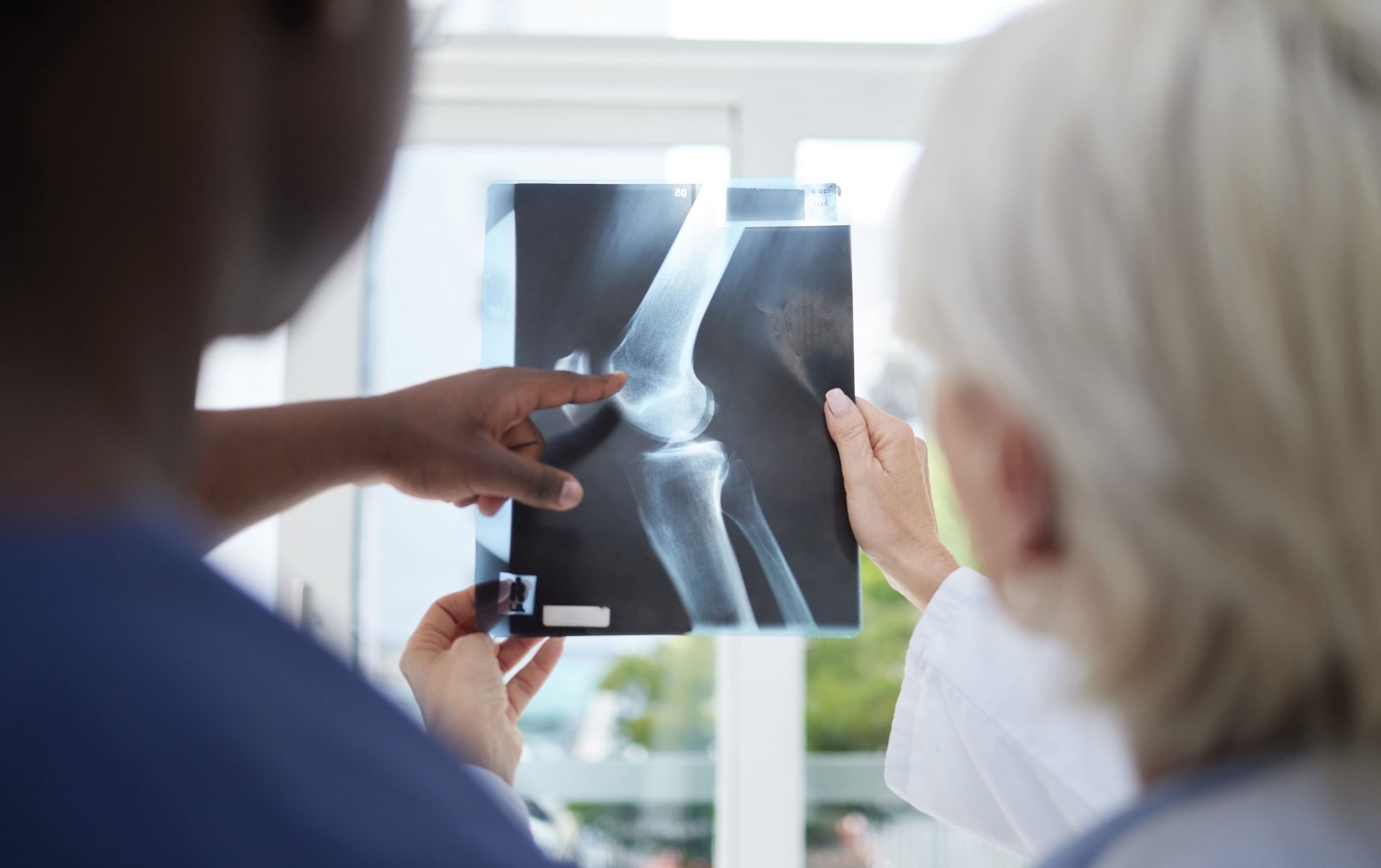
MRI (a scanning technique that uses magnets to create detailed images of the inside of your body) provides detailed visualisation of soft-tissue structures that cause mechanical symptoms. Modern sequences can detect meniscus tears, including subtle horizontal cleavage tears (cracks that run parallel to the meniscus surface) that may be missed on standard imaging. Cartilage sequences map areas of thinning or focal defects producing loose bodies (small fragments of tissue floating in the joint). Bone marrow oedema patterns (areas of swelling within the bone visible on scans) indicate areas of increased stress or early osteochondritis dissecans (a condition where a piece of bone and cartilage begins to separate). MRI arthrography with contrast injection (a special dye injected into the joint to enhance visualisation of structures) can improve the detection of labral tears (tears in the ring of cartilage around the hip or shoulder socket) and cartilage flaps.
Plain radiographs (X-rays) provide valuable information for detecting calcified loose bodies, advanced arthritis, and fractures. Standing weight-bearing views reveal joint space narrowing not apparent on images taken whilst lying down. Lateral views at various flexion angles may reveal loose bodies hidden in joint recesses. Merchant views (specific X-ray angles of the knee) assess patellofemoral alignment, which contributes to anterior knee locking. Sequential radiographs document the progression of degenerative changes correlating with worsening mechanical symptoms.
Diagnostic arthroscopy (a procedure in which a thin camera is inserted into the joint through a small incision) serves both diagnostic and therapeutic purposes when non-invasive imaging remains inconclusive. Direct visualisation can identify unstable cartilage flaps, partial thickness meniscus tears, and small loose bodies that may be missed on MRI. Dynamic assessment during arthroscopy reveals tissue impingement patterns during joint movement. Probing tests tissue stability and determines whether borderline lesions require treatment. Immediate treatment during diagnostic arthroscopy reduces the need for additional procedures, thereby shortening overall recovery time.
💡 Did You Know?
The knee joint contains mechanoreceptors (tiny sensors that detect movement and position) that detect joint position and movement. During locking episodes, these receptors send conflicting signals to your brain. Some indicate the joint should move whilst others detect mechanical obstruction. This creates the distinctive sensation of being “stuck” that differentiates true locking from simple stiffness.
Treatment Timelines
Treatment urgency depends on how often locking occurs, how severe it is, and what’s causing it. Actual mechanical locking (when you physically cannot fully straighten your knee) requires evaluation within days to prevent thigh muscle atrophy and joint stiffness. Locked bucket-handle meniscus tears (when a torn piece of cartilage flips into the joint) need arthroscopic surgery (a procedure using a small camera) within a short timeframe to minimise cartilage damage. Intermittent catching without actual locking allows scheduled evaluation and planned treatment.
Conservative management (non-surgical treatment) spans several weeks to months, depending on response. The initial phase focuses on reducing inflammation through activity modification and anti-inflammatory medications. Manual therapy techniques (such as joint mobilisation) may relieve minor mechanical blocks. Strengthening programmes require consistent effort over multiple weeks to support the joint. Progress markers include decreasing locking frequency, improved range of motion, and reduced pain with activities.
Surgical intervention timeline varies by procedure complexity. Simple arthroscopic loose-body removal (when the surgeon removes a small fragment using a camera and instruments) allows immediate weight-bearing and a return to normal activities within weeks. Meniscus repair requires protected weight-bearing for several weeks whilst tissue heals, followed by progressive rehabilitation over months. Complex reconstructive procedures (such as cartilage restoration or realignment surgery) demand extended recovery periods with specific milestone-based progression.
⚠️ Important Note
Forceful manipulation of a locked knee risks extending meniscus tears or displacing loose bodies into inaccessible joint regions. If your knee locks completely, try gentle rocking motions or gradual position changes. These often allow spontaneous unlocking. Persistent locking beyond several hours requires professional evaluation to prevent secondary damage.
Rehabilitation Strategies
Post-locking rehabilitation addresses both immediate symptoms and underlying mechanical dysfunction. Initial management reduces inflammation through ice application, compression, and elevation. Gentle range-of-motion exercises, such as slowly bending and straightening the knee, should be performed within pain-free limits. These help prevent adhesion formation (the sticking together of tissues). Isometric quadriceps contractions (tightening the thigh muscles without moving the joint) maintain muscle activation without stressing healing tissue. Patellar mobilisation techniques restore normal tracking that may contribute to locking episodes.
Progressive strengthening targets muscle imbalances that perpetuate abnormal joint mechanics. Quadriceps strengthening (exercises for the front thigh muscles) emphasises terminal extension (fully straightening the knee), which is important for preventing buckling episodes. Hamstring exercises (strengthening the back thigh muscles) balance anterior-posterior muscle forces. Hip strengthening, particularly of the gluteal muscles (buttock muscles), improves lower-extremity alignment, reducing abnormal knee stresses. Core stability work (exercises for abdominal and back muscles) enhances overall movement patterns during functional activities.
Neuromuscular training restores proprioception (your body’s sense of joint position and movement) disrupted by injury and locking episodes. Balance exercises progress from stable to unstable surfaces. Perturbation training (exercises that challenge your balance with unexpected pushes or movements) teaches automatic protective responses. Sport-specific drills incorporate cutting, pivoting, and deceleration movements, such as changing direction quickly, rotating on one foot, or stopping suddenly, that previously triggered locking. Biofeedback using mirrors or video can help correct movement patterns that contribute to mechanical symptoms.
Prevention Techniques for Active Individuals
- Implement progressive warm-up protocols incorporating dynamic stretching and gradual intensity increases before sports activities. Include knee-specific movements like leg swings, walking lunges, and controlled squats (exercises where you bend your knees in a controlled manner). These movements can help prepare joint structures for more demanding activities.
- Maintain a balanced strength ratio between the quadriceps (the muscles at the front of your thigh) and the hamstrings (the muscles at the back of your thigh) through targeted resistance training. Focus on eccentric strengthening exercises (movements in which you slowly lower the weight or resistance). These can help build tissue resilience against sudden deceleration forces that commonly cause meniscus tears.
- Practise landing mechanics during jumping activities by absorbing impact through hip and knee flexion (bending at your hips and knees) rather than stiff-legged landings. Train soft landings with bent knees. Engage the gluteal muscles (your buttocks) to help reduce direct knee stress.
- Modify training surfaces and equipment based on joint health status. Alternate between harder and softer running surfaces. Replace worn footwear before midsole compression becomes excessive (when the cushioning in your shoes becomes flattened). Adjust the bicycle seat height to maintain knee flexion angles.
- Schedule regular recovery periods to allow tissue adaptation between intense training sessions. Include active recovery activities like swimming or cycling. These maintain fitness whilst reducing repetitive knee stress patterns.
When to Seek Professional Help
- The knee remains stuck in a bent position for more than several hours despite gentle attempts to straighten it
- Repeated locking episodes occurring multiple times weekly or with increasing frequency
- Significant swelling develops shortly after locking episodes
- Inability to put weight on the affected leg after unlocking occurs
- Locking accompanied by sensations of knee instability, or the knee giving way during daily activities
- Progressive difficulty in straightening the knee fully, even when not actively locked
- Locking episodes following recent trauma or a fall onto the knee
- Night pain that may disrupt sleep patterns
- Visible knee deformity or abnormal joint shape during locked episodes
Commonly Asked Questions
What should I do immediately when my knee locks?
Avoid forceful attempts to unlock the knee. Sit or lie down to remove weight from the joint. Gently rock the knee using small movements. Apply ice to reduce pain and swelling. If the knee remains locked after an hour or you experience severe pain, seek medical evaluation. A gentle massage around the knee cap and surrounding muscles may help relax protective spasms (involuntary muscle contractions) that contribute to pseudo-locking.
Can knee locking be resolved without surgery?
Pseudo-locking from muscle spasms, mild arthritis, or minor inflammation can often improve with conservative treatment (non-surgical approaches such as physical therapy and activity modification). Actual mechanical locking from loose bodies (small fragments of bone or cartilage floating in the joint) or displaced meniscus tears (when the cartilage cushion in your knee shifts out of place) typically requires arthroscopic intervention (a procedure where a surgeon uses a small camera and instruments inserted through tiny incisions). Small, stable meniscus tears may heal without surgery if located in the vascular zone (the area of the meniscus with good blood supply). Your orthopaedic surgeon (a doctor who specialises in bones and joints) determines treatment based on imaging findings and clinical examination.
How long does recovery take after arthroscopic surgery for knee locking?
Recovery times vary depending on your specific procedure and individual health factors. Simple loose-body removal (removal of small fragments of bone or cartilage) allows normal walking within days and complete activities within a few weeks. Partial meniscectomy (removal of the damaged portion of the meniscus) requires gradual activity progression over several weeks. Meniscus repair (when the surgeon stitches the torn cartilage back together) demands protected weight-bearing for several weeks, followed by rehabilitation lasting several months. Individual factors, including age, pre-operative fitness, and rehabilitation compliance, affect recovery speed. Your surgeon can provide expectations for recovery based on your specific procedure and personal factors.
Will knee locking return after treatment?
Recurrence depends on the underlying cause and the completeness of treatment. Successful meniscus repair or complete removal of a loose body can often provide lasting relief. Degenerative conditions (conditions in which tissues gradually break down over time), such as arthritis, may produce new mechanical blocks. Maintaining strength and flexibility, and appropriately modifying activity, helps reduce the risk of recurrence. Regular follow-up identifies developing problems before significant mechanical symptoms return.
What activities should I avoid if I experience knee locking?
Avoid deep squatting, kneeling, and twisting movements that trigger locking episodes. High-impact activities (such as running or jumping) may worsen underlying damage. Modify problematic activities rather than complete avoidance when possible—use partial squats instead of deep squats, or swim instead of running. Your physiotherapist (a trained professional who specialises in movement and physical rehabilitation) can identify specific movement patterns causing symptoms and suggest appropriate modifications.
Conclusion
Mechanical locking due to a torn meniscus or loose bodies requires immediate evaluation, whilst pseudo-locking from inflammation may respond to conservative treatment. Proper diagnosis guides treatment selection between surgical and non-surgical approaches.
If you’re experiencing knee locking episodes, catching sensations, or sudden inability to straighten your knee, consult with an orthopaedic surgeon to evaluate your symptoms and determine appropriate treatment.

Experiencing Knee Pain or Injury?
Get a Personalised Treatment Plan
Find relief with our hip & knee specialists.
Make An Enquiry