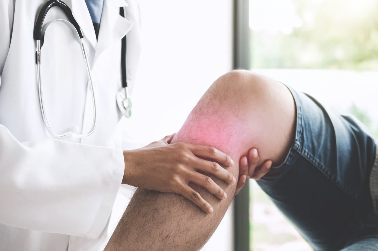
What happens when the ligament that keeps your knee stable during pivoting and cutting movements tears? The anterior cruciate ligament (ACL) stabilises your knee by preventing the tibia from sliding forward relative to the femur. When this ligament tears, the extent of damage determines the treatment approach and recovery timeline. Partial tears involve damage to only some ligament fibres, while complete tears result in total ligament disruption. MRI imaging reveals tears appearing as discontinuous fibres with increased signal intensity, though distinguishing between partial and complete tears often requires combining imaging with clinical examination findings.
ACL injuries occur through non-contact mechanisms in most cases – sudden pivoting, landing awkwardly from a jump, or rapid deceleration. Contact injuries also occur from direct blows to the knee. The distinction between partial and complete tears affects knee stability, treatment decisions, and return-to-activity timelines.
Structural Differences Between Tear Types
Complete ACL Tears
Complete tears involve total disruption of all ACL fibres. The ligament typically tears in the middle portion or avulses from its femoral or tibial attachment. On MRI, complete tears show absent ligament continuity with irregular fibre orientation. The torn ends often appear wavy or horizontally oriented rather than maintaining their normal diagonal course from femur to tibia.
Physical examination reveals positive Lachman test results with soft or absent endpoints. The anterior drawer test demonstrates increased tibial translation compared to the uninjured knee. Pivot shift testing, performed under anaesthesia, shows characteristic subluxation and reduction of the lateral tibial plateau.
Patients with complete tears experience immediate knee instability during pivoting activities. The knee may buckle or give way during direction changes, making participation in sports challenging without surgical reconstruction.
Partial ACL Tears
Partial tears preserve some intact ligament fibres while others sustain damage. These injuries affect either the anteromedial or posterolateral bundle, with anteromedial bundle tears occurring more frequently. MRI shows areas of increased signal intensity within the ligament alongside normal-appearing fibres.
Clinical examination findings vary based on the extent of the tear. Lachman testing may show increased laxity with a firm endpoint, distinguishing partial from complete tears. KT-1000 arthrometer measurements typically show side-to-side differences, whereas complete tears show greater differences.
Functional stability depends on which bundle remains intact and the percentage of fibres torn. Tears involving a smaller portion of ligament fibres often maintain adequate stability for daily activities, though high-demand sports may remain problematic.
Diagnostic Methods
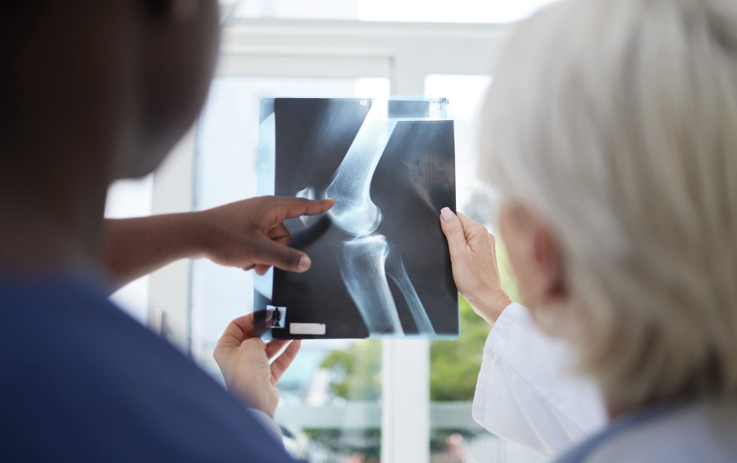
MRI Imaging
MRI is a commonly used imaging modality for evaluating ACL tears, with high accuracy for complete tears. Sagittal T2-weighted images demonstrate ligament continuity. Complete tears show absent ligament fibres or gross discontinuity. Partial tears display areas of increased signal intensity with some normal-appearing fibres remaining.
Secondary MRI signs support the diagnosis:
- Bone bruising patterns on the lateral femoral condyle and posterolateral tibial plateau
- Anterior tibial translation >7mm relative to the femur
- Buckling of the posterior cruciate ligament
- Deepening of the lateral femoral notch sign
Three-dimensional MRI sequences improve visualisation of individual ACL bundles, helping differentiate partial from complete tears when standard imaging remains inconclusive.
Clinical Examination Tests
The Lachman test provides sensitivity for ACL injuries. With the knee flexed 20-30 degrees, anterior tibial translation assessment reveals:
- Grade 1: <5mm translation with firm endpoint (often partial tear)
- Grade 2: 5-10mm translation with soft endpoint
- Grade 3: >10mm translation with absent endpoint (complete tear)
Anterior drawer testing at 90 degrees of flexion shows similar translation patterns but with lower sensitivity. The pivot shift test demonstrates rotational instability specific to complete ACL tears but requires patient relaxation for accurate assessment.
Instrumented laxity testing using KT-1000 or KT-2000 arthrometers provides objective measurements. Side-to-side differences >3mm at 30 pounds of force indicate ACL injury, with larger differences suggesting complete tears.
Treatment Approaches
Complete Tear Management
Complete ACL tears in active individuals typically require surgical reconstruction. Graft options include:
Bone-patellar tendon-bone (BPTB) autograft: Harvested from the central third of the patellar tendon with bone blocks from the patella and tibia. Provides strong initial fixation with bone-to-bone healing in tunnels within 6-8 weeks.
Hamstring tendon autograft: Semitendinosus and gracilis tendons provide a four-strand graft. Lower donor site morbidity compared to BPTB, but requires 12 weeks for tendon-to-bone healing.
Quadriceps tendon autograft: An option with clinical outcomes and minimal donor site complications. It can include a patellar bone block for hybrid fixation.
Non-operative management is suitable for sedentary individuals or those avoiding pivoting activities. Structured rehabilitation focuses on quadriceps and hamstring strengthening, proprioception training, and activity modification.
Partial Tear Treatment
Partial tears pose treatment challenges that require individualised approaches. Factors influencing management include:
- Tear extent (percentage of fibres involved)
- Bundle affected (anteromedial vs posterolateral)
- Patient activity level and sports demands
- Knee stability during functional testing
Non-operative treatment succeeds in many partial tears, particularly those involving less than half of the fibres. Rehabilitation emphasises:
- Immediate quadriceps activation exercises
- Progressive resistance training
- Neuromuscular control drills
- Sport-specific training for stable knees
Surgical options for partial tears include:
- Thermal shrinkage (abandoned mainly due to poor outcomes)
- Augmentation procedures preserving intact fibres
- Standard ACL reconstruction for progressive instability
⚠️ Important Note
Partial tears may progress to complete tears without proper rehabilitation or with premature return to pivoting sports.
Recovery Timelines
Complete Tear Rehabilitation
Post-reconstruction rehabilitation follows established protocols:
Weeks 0-2: Focus on reducing swelling, achieving full extension, and quadriceps activation. Weight bearing as tolerated with crutches.
Weeks 2-6: Progressive range of motion to 120 degrees flexion. Begin closed-chain strengthening exercises. Discontinue crutches when demonstrating a regular gait pattern.
Weeks 6-12: Full range of motion achievement. Advance strengthening program including leg press, squats, and hamstring curls. Initiate balance and proprioception exercises.
Months 3-6: Progressive strengthening with emphasis on single-leg exercises. Begin straight-line running at 4 months if strength on the involved side reaches appropriate levels compared to the uninvolved side.
Months 6-9: Sport-specific drills introduction. Agility training with progressive cutting activities. Isokinetic testing for appropriate strength symmetry.
Months 9-12: Return to sport consideration with functional testing. Psychological readiness assessment using ACL-RSI scale.
Partial Tear Recovery
Non-operative partial tear management typically allows faster return to activities:
Weeks 0-4: Emphasis on maintaining range of motion and reducing effusion. Immediate quadriceps strengthening within pain limits.
Weeks 4-8: Progressive strengthening program. Introduction of perturbation training for neuromuscular control.
Weeks 8-12: Progressive strengthening and functional exercises. Begin running the program if demonstrating reasonable control.
Months 3-4: Sport-specific training for stable knees. Return to sport decisions based on functional testing and absence of instability episodes.
Surgically treated partial tears follow similar timelines to complete reconstructions, though some surgeons allow accelerated protocols for augmentation procedures.
What Our Orthopaedic Surgeon Says
The distinction between partial and complete ACL tears significantly impacts treatment planning. While MRI provides valuable information, clinical examination findings and functional stability assessment are essential considerations. Many patients with partial tears maintain good function with proper rehabilitation, avoiding surgery altogether. However, young athletes in cutting sports often benefit from early reconstruction even with partial tears, as progressive instability can damage menisci and cartilage.
For complete tears, graft options are discussed based on patient factors – BPTB for young athletes requiring the fastest return to sport, hamstring grafts for recreational athletes concerned about kneeling pain, and quadriceps tendon for revision cases or larger individuals. Treatment is individualised based on tear pattern, patient goals, and functional demands rather than applying a one-size-fits-all approach.
Putting This Into Practice
- Request both MRI imaging and clinical examination for accurate tear classification, rather than relying on imaging alone.
- Document specific activities causing instability episodes to guide treatment decisions.
- Complete a full rehabilitation protocol regardless of tear type before returning to sports.
- Consider activity modification as a long-term strategy if choosing non-operative management for partial tears.
- Maintain quadriceps strength at 90% the uninjured side throughout recovery to protect the knee.
When to Seek Professional Help
- Immediate knee swelling after injury, limiting the range of motion
- Sensation of the knee “giving way” during daily activities
- Inability to bear weight on the affected leg
- Feeling of instability when changing direction
- Recurrent knee effusion after activities
- Conservative management has not led to improvement despite persistent instability.
- Mechanical symptoms suggesting an associated meniscal injury
Commonly Asked Questions
Can a partial ACL tear heal without surgery?
Partial ACL tears involving less than half of the fibres may scar to surrounding structures, providing functional stability. However, the ACL lacks robust blood supply, limiting healing potential. Success depends on compliance with rehabilitation and activity modification.
How accurate is MRI for determining the extent of a tear?
MRI is accurate for complete tears but less reliable for partial tears. Differentiating high-grade partial from complete tears remains challenging on imaging alone, necessitating correlation with clinical findings.
Will a partial tear eventually become complete?
Without proper rehabilitation and activity modification, partial tears risk progression. Continued pivoting activities with an unstable knee accelerate fibre damage. Structured rehabilitation reduces progression risk.
What factors determine surgery timing for partial tears?
Recurrent instability episodes, high activity demands, associated meniscal injury, and failure of conservative management indicate surgical intervention. Young athletes in cutting sports often benefit from early reconstruction.
How do outcomes differ between partial and complete tear treatments?
Appropriately treated partial tears often return to activities faster with less invasive treatment. Complete tears require longer rehabilitation but achieve predictable stability with reconstruction. Long-term outcomes depend more on rehabilitation quality than initial tear type.

Experiencing Knee Pain or Injury?
Get a Personalised Treatment Plan
Find relief with our hip & knee specialists.
Make An EnquiryConclusion
Accurate diagnosis, distinguishing partial from complete ACL tears, guides appropriate treatment selection. Complete tears typically require surgical reconstruction in active individuals, while partial tears may respond to conservative management. Both approaches demand comprehensive rehabilitation for optimal outcomes.
If you’re experiencing knee instability, giving way episodes, or recurrent swelling after activities, an orthopaedic surgeon can evaluate your condition and discuss treatment options.