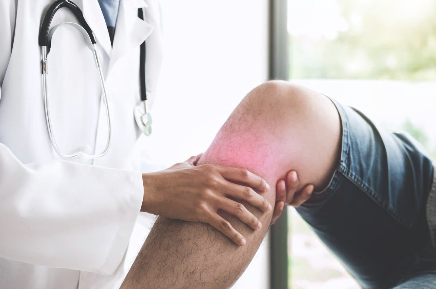
Does your knee hurt when you walk up stairs, and you’re not sure why? Patellofemoral pain syndrome (PFPS) causes pain around and behind the kneecap, particularly during activities like running, squatting, or climbing stairs. The condition occurs when increased stress between the kneecap (patella) and thighbone (femur) leads to irritation of the joint surfaces. Unlike other knee injuries that involve torn ligaments or damaged cartilage, PFPS arises from biomechanical issues that alter the way the kneecap tracks in its groove.
The pain typically starts gradually and worsens with continued activity. Many people first notice discomfort during or after exercise, especially activities involving repetitive knee bending. The condition affects both recreational and competitive athletes, though anyone who suddenly increases their activity level may develop symptoms.
Anatomy of the Patellofemoral Joint
The patellofemoral joint consists of the kneecap (patella) and the groove at the end of the thighbone (femoral trochlea) where it sits. The patella acts as a fulcrum, increasing the leverage of the quadriceps muscle during knee extension. A layer of cartilage covers both the back of the patella and the femoral groove, allowing smooth gliding motion during movement.
Four main structures control patellar movement and stability:
- The quadriceps muscle group provides the primary force for knee extension while also controlling patellar position
- The patellar tendon connects the kneecap to the shinbone, transmitting force during movement
- Medial and lateral retinaculae – fibrous tissues on either side of the patella – help guide tracking
- The iliotibial (IT) band, running along the outer thigh, can influence lateral patellar movement when tight
Normal patellar tracking requires balanced forces from all surrounding structures. During knee flexion, the patella glides downward in the femoral groove. At full extension, it sits slightly above and to the side of the groove. Any disruption to this movement pattern—whether from muscle weakness, tightness, or structural variations—can lead to abnormal stress and pain.
Causes and Risk Factors
Muscle imbalances represent a common cause of PFPS. Weakness in the hip abductors and external rotators allows the knee to collapse inward during weight-bearing activities, altering patellar tracking. Specifically, gluteus medius weakness fails to control femoral rotation, while vastus medialis oblique (VMO) weakness reduces medial patellar stability. This combination creates lateral patellar drift and increased contact pressure.
Training errors frequently trigger PFPS onset. Sudden increases in running distance, intensity, or frequency overwhelm the body’s adaptation capacity. Changes in running surface—transitioning from soft trails to concrete or from flat terrain to hills—alter loading patterns. Inadequate recovery between training sessions prevents tissue adaptation and repair.
Biomechanical factors contribute significantly to the development of PFPS. Excessive foot pronation increases internal tibial rotation, affecting patellar alignment. Hip internal rotation and adduction during single-leg stance activities create dynamic valgus collapse. Reduced ankle dorsiflexion forces compensatory movements at the knee. These patterns often combine, creating cumulative stress on the patellofemoral joint.
💡 Did You Know?
The patella experiences significant forces during activities like squatting or jumping, explaining why these movements often trigger symptoms in PFPS.
Recognizing Symptoms
PFPS pain typically localises to the front of the knee, either around or behind the kneecap. Patients often describe a dull, aching sensation rather than sharp pain. The discomfort usually develops gradually over weeks or months, though some people report a sudden onset after a particularly intense training session.
Specific activities consistently aggravate PFPS symptoms:
- Prolonged sitting with bent knees causes “theatre sign” – pain and stiffness requiring frequent position changes
- Descending stairs typically hurts more than ascending due to higher patellofemoral joint forces
- Squatting, kneeling, and running (especially downhill) commonly trigger symptoms
- Many patients report a grinding or clicking sensation during knee movement, though this doesn’t always correlate with pain severity.
Morning stiffness that improves with movement characterises many PFPS cases. The knee may feel unstable or weak, particularly during eccentric muscle contractions, such as lowering into a squat. Some patients experience mild swelling around the kneecap, though significant effusion suggests other pathology. Symptoms often affect both knees, though one side usually predominates.
Diagnosis Process
Clinical examination forms the foundation of PFPS diagnosis. Orthopaedic surgeons assess patellar mobility, checking for excessive lateral translation or tilt. The patellar compression test—pressing the kneecap against the femur while the patient contracts their quadriceps—typically reproduces symptoms. Palpation along the patellar borders often reveals tenderness, particularly at the lateral facet.
Functional movement assessment reveals biomechanical contributors. Single-leg squat testing demonstrates dynamic knee valgus, hip drop, or trunk lean. Step-down tests show similar patterns as eccentric control demands are added. Flexibility testing identifies tight structures, including the hip flexors, quadriceps, IT band, and calf muscles. Strength testing focuses on hip abductors, external rotators, and quadriceps, comparing side-to-side differences.
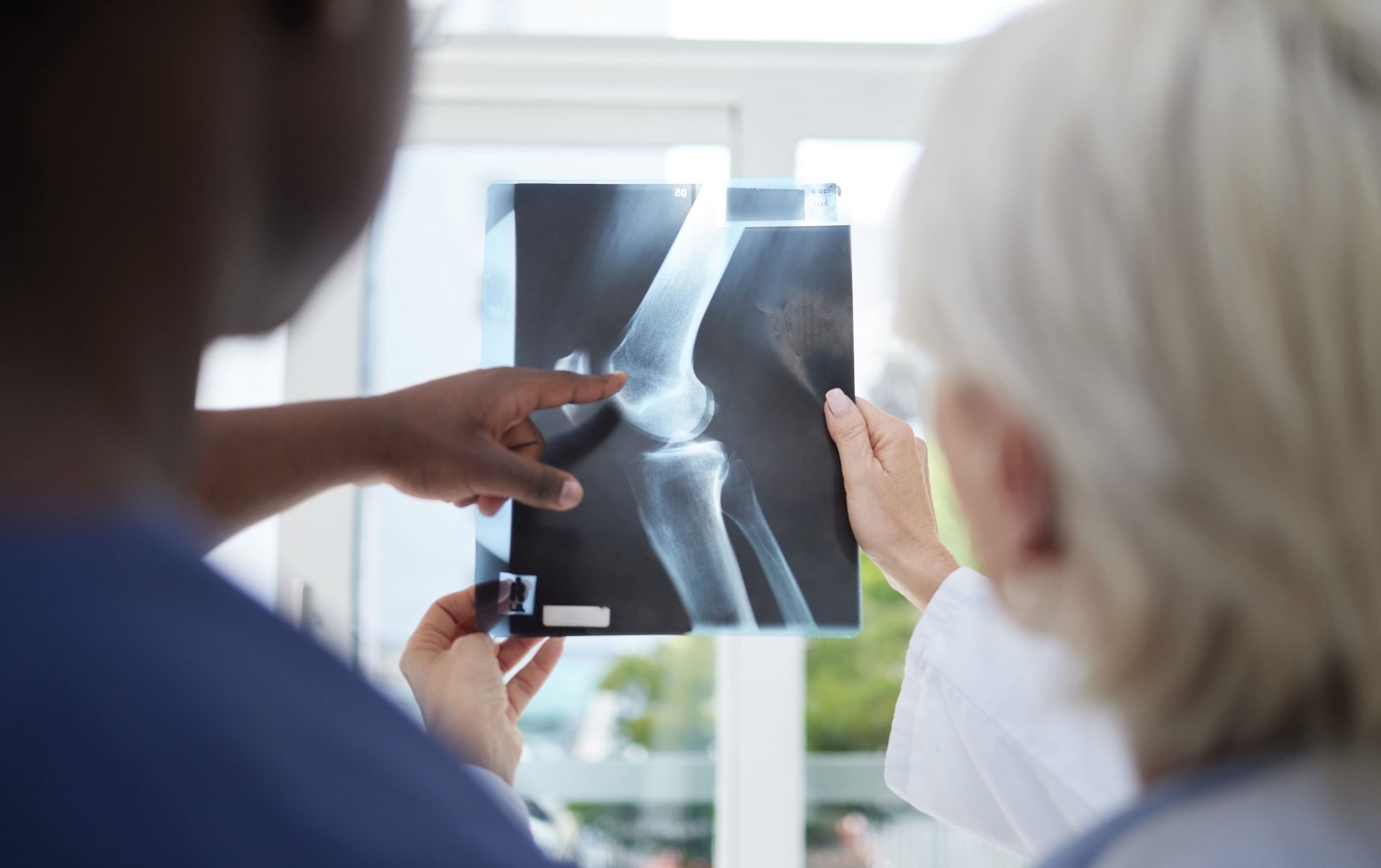
Imaging rarely changes PFPS management but may rule out other conditions. X-rays can show patellar tilt, subluxation, or arthritic changes. MRI reveals cartilage abnormalities, bone oedema, or soft-tissue abnormalities. However, imaging findings don’t always correlate with symptoms—some people have abnormal findings without pain, while others have normal findings despite significant symptoms.
⚠️ Important Note
Red flags requiring immediate evaluation include significant swelling, inability to bear weight, knee locking or giving way, fever, or pain unrelated to activity – these suggest conditions beyond PFPS.
Conservative Treatment Approaches
Activity modification initiates the healing process without requiring complete rest. Reducing training volume allows continued participation while decreasing symptoms. Substituting high-impact activities with cycling or swimming maintains fitness while reducing patellofemoral stress. Avoid prolonged sitting with bent knees, and use a small pillow under the knee when sitting to help manage daily symptoms.
Targeted strengthening addresses the muscle imbalances associated with PFPS. Hip strengthening begins with side-lying abduction, progressing to standing exercises with resistance bands. Clamshells and hip external rotation exercises target deep hip stabilisers. Quadriceps strengthening starts with straight-leg raises and wall sits, progressing to single-leg squats and step-downs as pain allows. Eccentric exercises – slowly lowering into positions – build control and strength simultaneously.
Manual therapy techniques complement exercise programs. Patellar mobilisation improves tracking and reduces stiffness. Soft-tissue work addresses tight lateral structures that pull the patella off course. IT band stretching and foam rolling, combined with hip flexor and calf stretching, restore standard movement patterns. Taping techniques can provide immediate pain relief by improving patellar position during activities.
✅ Quick Tip
Ice application for 15-20 minutes after activity reduces inflammation. Elevating the leg above heart level during icing may enhance effectiveness by promoting fluid drainage.
Running Modifications
Gait analysis reveals running patterns contributing to PFPS:
- Overstriding – landing with the foot far ahead of the body’s centre of mass – increases braking forces and patellofemoral stress
- A cadence below 170 steps per minute often correlates with overstriding
- Excessive vertical oscillation wastes energy and increases impact forces
- Hip drop during the stance phase indicates weak hip stabilisers unable to control pelvic position
Running technique adjustments can reduce symptoms:
- Increasing cadence by 5-10% shortens stride length and reduces impact forces
- Landing with a slight forward trunk lean shifts the load away from the patellofemoral joint
- Focusing on “quiet” foot strikes minimises impact
- Maintaining level hips through conscious glute engagement improves alignment
These changes require gradual implementation—altering too many variables simultaneously often creates new problems.
Progressive return to running follows symptom resolution. Initial sessions combine walking and jogging intervals on flat, soft surfaces. Duration increases before intensity, with pain monitoring guiding progression. Hill running resumes last due to higher patellofemoral forces. Cross-training maintains fitness during the return phase, with cycling particularly useful as it strengthens quadriceps without impact.
What Our Orthopaedic Surgeon Says
PFPS recovery requires patience and consistency. Addressing the underlying biomechanical issues takes time. Some discomfort during rehabilitation is acceptable—typically 2-3 out of 10, resolving within 24 hours. This approach allows progressive loading while respecting tissue healing.
Returning to full activity too quickly is a common mistake. Just because symptoms improve doesn’t mean the underlying issues are resolved. Continuing strengthening exercises for several months after symptoms resolve prevents recurrence. Periodic movement assessments can help catch problems before they become symptomatic again.
Treatment Options
Persistent symptoms despite conservative treatment may warrant additional interventions. Corticosteroid injections can provide temporary relief for inflammatory components, though they don’t address underlying mechanical issues. Platelet-rich plasma (PRP) injections show promise for stimulating tissue healing, particularly in cases involving cartilage. Hyaluronic acid injections may improve joint lubrication and reduce pain.
Physical therapy modalities supplement exercise programs. Neuromuscular electrical stimulation helps activate weak muscles, particularly the VMO. Biofeedback training improves movement patterns by providing real-time information about muscle activation. Blood flow restriction training allows strength gains with lighter loads, useful when heavy resistance aggravates symptoms.
Surgical intervention rarely becomes necessary for PFPS. Lateral release procedures address excessive lateral patellar tilt by cutting tight retinacular tissues. Tibial tubercle transfer repositions the patellar tendon attachment to improve tracking. Cartilage restoration procedures address focal defects contributing to symptoms. However, surgery addresses only structural issues; biomechanical factors still require correction through rehabilitation.
Long-term Management Strategies
Preventing PFPS recurrence requires ongoing attention to contributing factors. Maintenance strengthening programs focus on hip and core stability and are performed 2-3 times weekly, even when asymptomatic. Regular flexibility work addresses areas prone to tightness. Gradual training progressions respect tissue adaptation timelines.
Equipment considerations play a supporting role. Running shoes with appropriate cushioning and support for individual foot types reduce impact forces. Replacing shoes regularly prevents breakdown of midsole materials. Patellar straps or sleeves provide proprioceptive feedback and mild support during activities. Custom orthotics address biomechanical issues identified during assessment.
Monitoring early warning signs allows prompt intervention. Mild anterior knee discomfort after activity signals the need for training modification. Increased morning stiffness suggests inadequate recovery. Regular self-assessment using single-leg squat tests identifies returning movement dysfunction. Addressing these signs immediately prevents progression to limiting symptoms.
Commonly Asked Questions
How long does PFPS typically take to resolve?
Recovery timelines vary based on symptom duration and contributing factors. Acute cases often improve within 6-12 weeks of appropriate treatment. Chronic cases may require 3-6 months of consistent rehabilitation. Complete resolution includes not just pain relief but also correction of underlying biomechanical issues to prevent recurrence.
Can I continue exercising with PFPS?
Modified activity usually proves beneficial rather than complete rest. Low-impact activities like cycling, swimming, or elliptical training maintain fitness while allowing recovery. The approach involves identifying activities that don’t increase symptoms beyond mild discomfort, which resolves within 24 hours. Gradual return to impact activities follows initial symptom improvement.
Why does sitting make my knee hurt?
Prolonged knee flexion increases patellofemoral contact pressure. Sitting positions the patella deep within the femoral groove under tension from the quadriceps. This sustained pressure irritates already sensitive tissues. Regular position changes, using a footrest to reduce knee flexion, or placing a small pillow behind the knee, help manage sitting-related symptoms.
Should I use knee supports or braces?
Patellar stabilising braces or sleeves can provide symptom relief during activities. They work through proprioceptive feedback and mild mechanical support rather than correcting patellar position. While useful for symptom management, braces shouldn’t replace addressing underlying strength and flexibility deficits. Braces can be helpful during the rehabilitation process, but may become unnecessary once recovered.
When is surgery necessary for PFPS?
Surgical intervention becomes an option only after conservative treatment for 6-12 months has not provided adequate improvement. Specific indications include documented patellar instability, significant cartilage damage, or structural abnormalities preventing normal tracking. Surgery addresses structural issues but still requires extensive rehabilitation to correct movement patterns.

Experiencing Knee Pain or Injury?
Get a Personalised Treatment Plan
Find relief with our hip & knee specialists.
Make An EnquiryConclusion
Effective PFPS management may require addressing both symptoms and underlying biomechanical causes by targeting the strengthening of hip and knee stabilisers. Most patients achieve significant improvement with consistent rehabilitation focusing on movement pattern correction and gradual activity progression.
If you’re experiencing persistent knee pain around the kneecap, difficulty with stairs or squatting, or pain during running activities, consultation with an orthopaedic surgeon can provide a comprehensive evaluation and specialised treatment options.