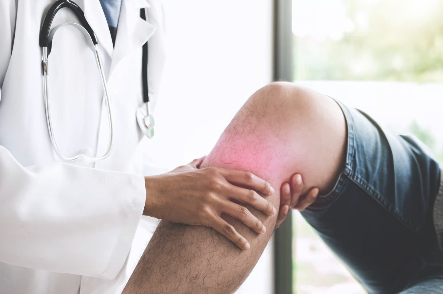
Did you know that a single misstep can tear multiple knee ligaments simultaneously, creating complex instability patterns that affect your ability to walk normally? The knee contains four primary ligaments that stabilise the joint during movement. Each ligament serves a specific function, and tears to these structures create distinct patterns of instability and symptoms.
Ligament tears range from minor stretching (Grade 1) to complete ruptures (Grade 3). The mechanism of injury, pain location, and specific instability pattern often indicate which ligament was injured before imaging confirms the diagnosis.
Anterior Cruciate Ligament (ACL) Tears
ACL tears typically occur during sudden direction changes, jumping, or direct impact to the knee. The ligament prevents the tibia from sliding forward relative to the femur and provides rotational stability during pivoting movements.
Injury Mechanism and Symptoms
ACL injuries often happen without contact – landing awkwardly from a jump, rapidly changing direction, or suddenly stopping while running. Many patients report hearing a “pop” at the time of injury, followed by immediate swelling within 2-6 hours.
The knee typically feels unstable during twisting movements or when walking on uneven surfaces. Patients describe the sensation as the knee “giving way” or feeling like it might buckle. Full knee extension often becomes difficult due to swelling and pain.
Diagnosis Methods
Physical examination includes the Lachman test, in which the orthopaedic surgeon assesses excessive forward movement of the tibia. The anterior drawer test and pivot shift test provide additional assessment of ligament integrity.
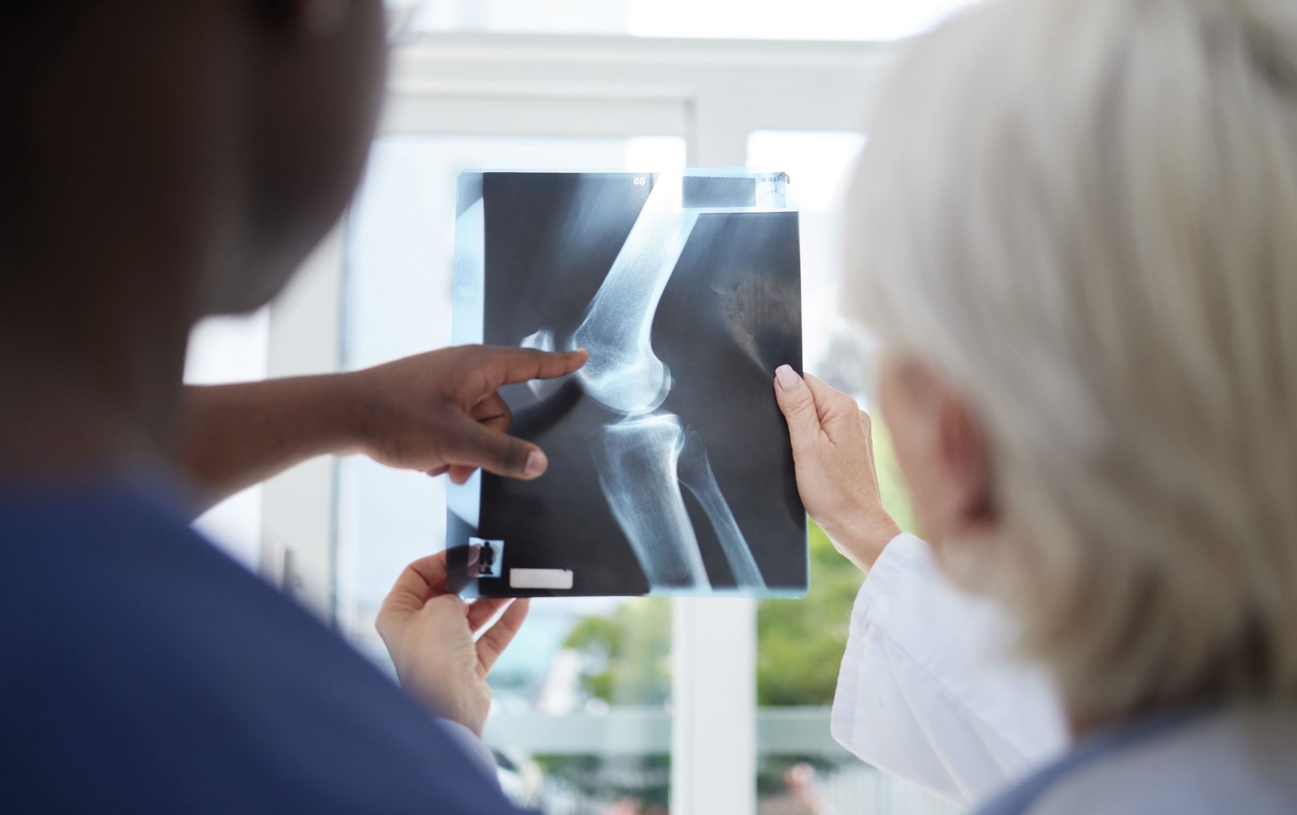
MRI scanning confirms the diagnosis and reveals associated injuries such as meniscal tears or bone bruising. The imaging shows disruption of the ligament fibres and helps determine if the tear is partial or complete.
Treatment Approaches
Non-surgical management is suitable for patients with partial tears or lower activity demands. This involves:
- Structured physiotherapy focusing on quadriceps and hamstring strengthening
- Proprioception exercises to improve joint position awareness
- Activity modification, avoiding pivoting in sports
- Functional knee bracing during activities
Surgical reconstruction becomes necessary for complete tears in active individuals or those experiencing persistent instability. The procedure involves replacing the torn ACL with a hamstring, patellar tendon, or quadriceps tendon graft. Recovery typically requires 9-12 months before returning to pivoting sports.
Posterior Cruciate Ligament (PCL) Tears
PCL injuries occur less frequently than ACL tears and usually result from direct trauma to the front of the knee. This ligament prevents backward movement of the tibia and works with the ACL to control rotation.
Common Causes
Dashboard injuries during car accidents represent the typical PCL injury mechanism – the bent knee strikes the dashboard, forcing the tibia backwards. Sports-related PCL tears happen when athletes fall onto a flexed knee with the foot pointed downward.
Unlike ACL injuries, PCL tears rarely occur in isolation. Associated injuries include damage to the posterolateral corner structures or other ligaments.
Clinical Presentation
PCL tears produce less dramatic symptoms compared to ACL injuries. Swelling develops gradually over several hours or days rather than immediately. Patients report difficulty with deceleration activities, such as walking down stairs or downhill.
The posterior sag sign becomes evident when the knee is examined at 90 degrees of flexion: the affected tibia sits further back than the uninjured side. Pain localises to the back of the knee and increases with deep knee bending.
Management Strategies
Partial PCL tears and isolated complete tears often heal without surgery due to the ligament’s good blood supply. Conservative treatment includes:
- Quadriceps strengthening exercises starting immediately
- Progressive weight-bearing as tolerated
- Avoiding hamstring exercises initially to prevent posterior tibial translation
- Gradual return to activities over 3-4 months
Surgical reconstruction addresses combined ligament injuries or failed conservative treatment. The timing of surgery depends on associated injuries and the patient’s functional demands.
Medial Collateral Ligament (MCL) Tears
The MCL runs along the inner side of the knee, connecting the femur to the tibia. This ligament resists forces that push the knee inward and provides stability during normal walking and running.
Injury Patterns
MCL tears result from blows to the outer side of the knee, forcing it inward. Contact sports produce MCL injuries through tackles or collisions. The ligament can also tear during twisting injuries that stress the inner knee.
Tears occur at three locations: the femoral attachment (most common), mid-substance, or tibial attachment. The location affects healing potential and treatment decisions.
Symptoms and Examination
Pain and tenderness concentrate along the inner knee, with swelling developing over the medial joint line. Patients experience pain when the knee bends beyond 30 degrees or during activities that stress the inner knee.
The valgus stress test evaluates MCL integrity by applying outward pressure to the knee at 0 and 30 degrees of flexion. Increased joint opening compared to the uninjured side indicates ligament damage. The degree of opening correlates with injury severity.
Recovery Process
MCL tears have a good healing potential due to their blood supply. Treatment typically involves:
- Hinged knee bracing limiting valgus stress for 4-6 weeks
- Early range of motion exercises within pain limits
- Progressive strengthening focusing on the quadriceps and hip muscles
- Sport-specific rehabilitation before return to activities
Grade 3 MCL tears with associated injuries may require surgical repair or reconstruction. Chronic MCL insufficiency, which may lead to persistent instability, may also warrant surgical intervention.
Lateral Collateral Ligament (LCL) Tears
LCL injuries represent the least common isolated knee ligament tear. The LCL connects the femur to the fibula on the outer knee and works with surrounding structures to prevent excessive outward knee movement.
Mechanism and Associated Injuries
Direct blows to the inner knee force it outward, stressing the LCL. Hyperextension injuries can also damage this ligament. Isolated LCL tears rarely occur – most involve the posterolateral corner structures, including the popliteus tendon and popliteofibular ligament.
Combined injuries create rotational instability in addition to lateral instability, significantly affecting knee function.
Clinical Features
Patients report lateral knee pain with point tenderness at the fibular head. Swelling remains minimal compared to other ligament injuries. Walking feels unstable, particularly on uneven surfaces or when changing direction.
The varus stress test checks LCL integrity by pushing the knee inward. Increased lateral joint opening indicates LCL damage. The dial test and posterolateral drawer test evaluate associated posterolateral corner injuries.
Treatment Considerations
Isolated partial LCL tears may respond to conservative management:
- Protected weight-bearing with crutches initially
- Knee bracing prevents varus stress
- Strengthening exercises for hip abductors and lateral thigh muscles
- Balance and proprioception training
Complete tears or combined posterolateral corner injuries typically require surgical reconstruction. Delay in treating these complex injuries leads to progressive instability and early arthritis. Surgery aims to restore both static and dynamic knee stability.
Multi-Ligament Knee Injuries
Severe knee trauma can damage multiple ligaments simultaneously, creating complex instability patterns. These injuries result from high-energy mechanisms such as motor vehicle accidents, falls from height, or severe sports collisions.
Classification and Assessment
Multi-ligament injuries involve two or more major ligaments. The most severe pattern, knee dislocation, damages at least three ligaments and risks neurovascular injury. Even if the knee relocates spontaneously, the underlying ligament damage remains.
Immediate evaluation includes checking blood flow to the foot and nerve function. The ankle-brachial index measurement screens for arterial injury requiring urgent vascular surgery consultation.
Surgical Planning
Multi-ligament reconstructions require careful timing and sequencing. Acute surgery within three weeks addresses specific injury patterns, while others benefit from staged procedures after swelling resolves.
Surgeons may restore proper knee kinematics by addressing each damaged structure. This may involve:
- Ligament repairs for avulsion injuries
- Reconstructions using multiple grafts
- Addressing associated cartilage or meniscal damage
- Correcting limb alignment if necessary
Rehabilitation Challenges
Recovery from multi-ligament surgery extends beyond single ligament timelines. Patients require extended recovery periods before returning to high-demand activities. The rehabilitation program balances protecting healing tissues while preventing stiffness and muscle atrophy.
Close monitoring may allow appropriate progression through rehabilitation phases. Functional testing guides return-to-activity decisions rather than relying solely on time-based protocols.
Putting This Into Practice
- Document the injury mechanism, including body position and forces involved, to help your orthopaedic surgeon identify likely injured structures.
- Apply ice and compression immediately after injury, elevating the leg above heart level to minimise swelling.
- Use crutches to avoid weight-bearing until examined by a medical professional to prevent further damage to the injured ligaments.
- Begin gentle quadriceps contractions while waiting for medical evaluation to maintain muscle activation.
- Keep a symptom diary, noting specific activities that cause instability or pain to guide treatment planning.
When to Seek Professional Help
- Immediate inability to bear weight on the injured leg
- Visible knee deformity or abnormal leg alignment
- Rapid swelling within two hours of injury
- Feeling or hearing a “pop” during the injury
- Knee buckling or giving way during normal activities
- Persistent pain along the joint line after several days
- Inability to fully straighten or bend the knee
- Numbness or tingling below the knee
- Cold foot or absent pulse below the injury
Commonly Asked Questions
How long will it take before I can return to sports after ligament surgery?
Return timelines vary by ligament and sport demands. ACL reconstruction typically requires 9-12 months before returning to cutting sports. MCL injuries may allow return within 6-8 weeks for grade 1-2 tears. An orthopaedic surgeon determines clearance based on strength testing, functional assessments, and sport-specific demands rather than time alone.
Can knee ligaments heal without surgery?
MCL and partial PCL tears often heal without surgery due to good blood supply. Complete ACL tears in adults cannot heal independently due to poor blood supply and the synovial environment. Treatment decisions depend on tear severity, patient age, activity level, and associated injuries.
What determines whether I need immediate surgery or can wait?
Multi-ligament injuries with documented vascular compromise require emergency surgery. Most isolated ligament tears allow time for swelling reduction before surgery. Locked knees from associated meniscal tears may need urgent arthroscopy. An orthopaedic surgeon evaluates injury patterns, associated damage, and tissue quality when determining the timing of surgery.
Will I develop arthritis after a ligament tear?
Ligament injuries increase the risk of arthritis, particularly when associated with cartilage damage or chronic instability. Proper treatment and rehabilitation minimise but cannot eliminate this risk. Maintaining a healthy weight, making appropriate activity modifications, and engaging in regular strengthening exercises help protect the knee in the long term.
How do I know if my knee instability is due to ligament damage or muscle weakness?
Ligament instability creates specific directional giving way – forward with ACL tears, inward with MCL injuries. Muscle weakness causes general knee instability without directional patterns. Physical examination by an orthopaedic surgeon differentiates between structural and functional instability.

Experiencing Knee Pain or Injury?
Get a Personalised Treatment Plan
Find relief with our hip & knee specialists.
Make An EnquiryConclusion
Accurate diagnosis determines whether your ligament tear requires surgery or conservative management. Early evaluation prevents secondary damage from chronic instability and guides appropriate rehabilitation. Treatment success depends on identifying the specific ligaments involved and promptly addressing associated injuries.
If you’re experiencing knee instability, buckling, or persistent pain after injury, an orthopaedic surgeon can evaluate your condition and discuss treatment options.