Can your knee bend backwards far enough to tear the ligaments inside? Knee hyperextension occurs when the knee joint bends backwards beyond its normal 0-degree straight position, sometimes reaching substantial posterior angulation. This movement places stress on the ligaments, joint capsule, and surrounding structures of the posterior knee. The injury ranges from mild ligament stretching to complete tears of stabilising structures, depending on the force and direction of impact. Athletes in jumping and pivoting sports frequently encounter this injury. It also occurs during everyday activities, such as missing a step or landing awkwardly from a height.
The knee joint relies on four primary ligaments to maintain stability throughout its range of motion:
- The anterior cruciate ligament (ACL, which prevents the shin bone from sliding too far forward.
- The posterior cruciate ligament (PCL, which prevents the shin bone from sliding backwards.
- The medial and collateral ligaments
Hyperextension primarily affects the ACL and PCL, as these structures resist forward-backwards movement of the tibia (shin bone) relative to the femur (thigh bone).
How Knee Hyperextension Happens
The knee hyperextends when a straightening force continues beyond the point at which the joint normally stops. Landing from a jump with the knee locked straight creates a common mechanism. Ground reaction force drives the tibia backwards relative to the femur. Direct blows to the front of the knee, with the foot fixed, produce similar hyperextension forces.
Sports involving sudden deceleration carry particular risk. A footballer planting their foot to change direction, whilst another player contacts the front of their thigh, experiences combined hyperextension and rotational stress. Basketball players landing from rebounds with locked knees face pure hyperextension forces multiplied by body weight and downward momentum.
Non-sporting causes include stepping into unexpected holes, missing stairs, and motor-vehicle accidents in which the knee strikes the dashboard. These mechanisms often involve greater forces and less neuromuscular preparation than those in athletic injuries.
Anatomical Vulnerability
The posterior joint capsule (the fibrous tissue that surrounds the back of the knee joint) provides the first line of resistance against hyperextension. When stretched beyond its capacity, the capsule tears, allowing further backward movement. The ACL then engages as the primary restraint. The PCL follows if forces continue.
The popliteal artery (the blood vessel that runs directly behind the knee joint) makes severe hyperextension injuries potentially serious. Arterial damage remains uncommon in typical sports injuries. High-energy trauma requires urgent vascular assessment.
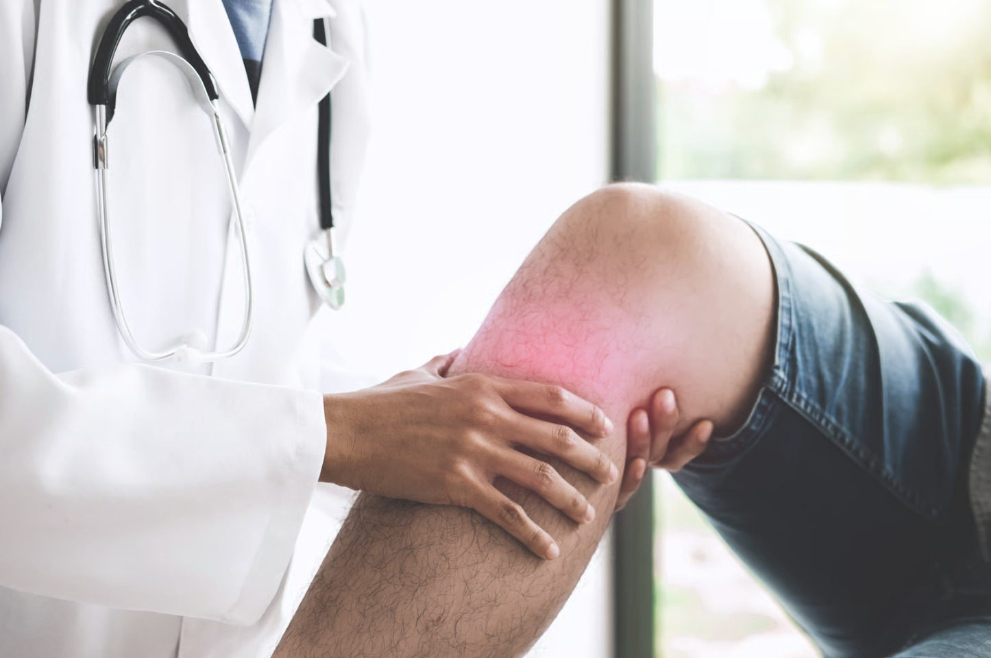
Recognising Hyperextension Symptoms
Immediate symptoms following knee hyperextension include a sensation of the knee “giving way” backwards, often described as the joint bending in the wrong direction. Pain initially localises to the back of the knee. Anterior knee pain develops as swelling increases within hours.
Swelling patterns help differentiate injury severity. Rapid swelling within the first two hours suggests bleeding into the joint (haemarthrosis, meaning blood has collected within the joint space). This indicates likely ligament damage. Gradual swelling over 24-48 hours typically reflects synovial fluid accumulation (an increase in the joint’s natural lubricating fluid) from capsular irritation rather than structural tearing.
Weight-bearing ability varies considerably. Some individuals walk immediately after hyperextension injuries that later prove to involve complete ACL tears. The surrounding muscles temporarily compensate for ligament laxity. Others cannot bear weight after minor sprains due to pain and protective muscle spasm.
Instability sensations during subsequent activities provide diagnostic information. Feelings of the knee “shifting” during pivoting movements suggest ligament insufficiency. Difficulty fully straightening the knee may indicate swelling, muscle guarding, or mechanical blocking due to an associated meniscal injury.
💡 Did You Know?
The knee’s normal extension stops at 0 degrees in most individuals, though some people naturally hyperextend several degrees without injury. This physiological hyperextension, called genu recurvatum, results from ligament laxity and requires greater muscular control during activities.
Structures Commonly Affected
Anterior Cruciate Ligament
The ACL is commonly injured in hyperextension injuries, particularly those involving rotational components. Complete tears produce immediate instability. Initial swelling and muscle guarding may mask this finding. Partial tears create variable symptoms depending on the percentage of fibres disrupted and the individual’s activity demands.
Posterior Cruciate Ligament
Pure hyperextension mechanisms place substantial stress on the PCL. Isolated PCL injuries more commonly result from dashboard impacts during vehicle collisions. Combined ACL-PCL injuries occur with extreme hyperextension forces. These create complex instability patterns requiring careful surgical planning.
Posterior Joint Capsule
Capsular stretching or tearing accompanies most hyperextension injuries. The capsule heals by forming scar tissue. Residual laxity may persist. Capsular injuries alone rarely cause functional instability but contribute to the overall assessment of injury severity.
Meniscal Involvement
The menisci—crescent-shaped cartilage structures cushioning the joint—may tear during hyperextension, particularly when combined with rotation. Posterior horn tears commonly accompany ACL injuries. Symptoms include mechanical catching and locking, and localised joint-line tenderness distinct from ligament-related pain.
Initial Management Following Injury
Immediate management focuses on controlling swelling and preventing further damage. The PRICE protocol—Protection, Rest, Ice, Compression, Elevation—remains standard initial care. Apply ice for 15-20 minutes every 2-3 hours during waking hours to help limit the inflammatory response.
Protected weight-bearing with crutches reduces stress on damaged structures. Full weight-bearing restriction typically continues until comfortable walking becomes possible, usually within days for minor injuries. Knee braces or immobilisers provide external stability during the acute phase. Prolonged immobilisation leads to muscle weakness and joint stiffness.
Begin range-of-motion exercises once acute pain subsides. Gentle bending and straightening within comfortable limits prevent adhesion formation (scar tissue sticking together) and maintain cartilage nutrition. Quadriceps setting exercises—tightening the thigh muscle without joint movement—preserve muscle activation patterns even when movement remains limited.
⚠️ Important Note
Inability to bear any weight, severe swelling within two hours of injury, obvious deformity, or numbness below the knee requires urgent medical evaluation. These findings suggest potentially serious structural damage or neurovascular compromise (disruption to nerves or blood vessels).
Diagnostic Evaluation Process
Clinical examination begins with observation of swelling, bruising, and resting knee position. Palpation (gentle pressure and tactile assessment of the area) identifies areas of maximal tenderness. This helps localise injured structures. Specific ligament tests assess stability in multiple directions.
The Lachman test (a clinical assessment in which the physician moves the tibia forward while stabilising the femur) evaluates ACL integrity by assessing tibial translation with the knee slightly bent. The posterior drawer test assesses PCL function by measuring posterior tibial displacement. Valgus and varus stress testing check medial and lateral collateral ligament stability, respectively.
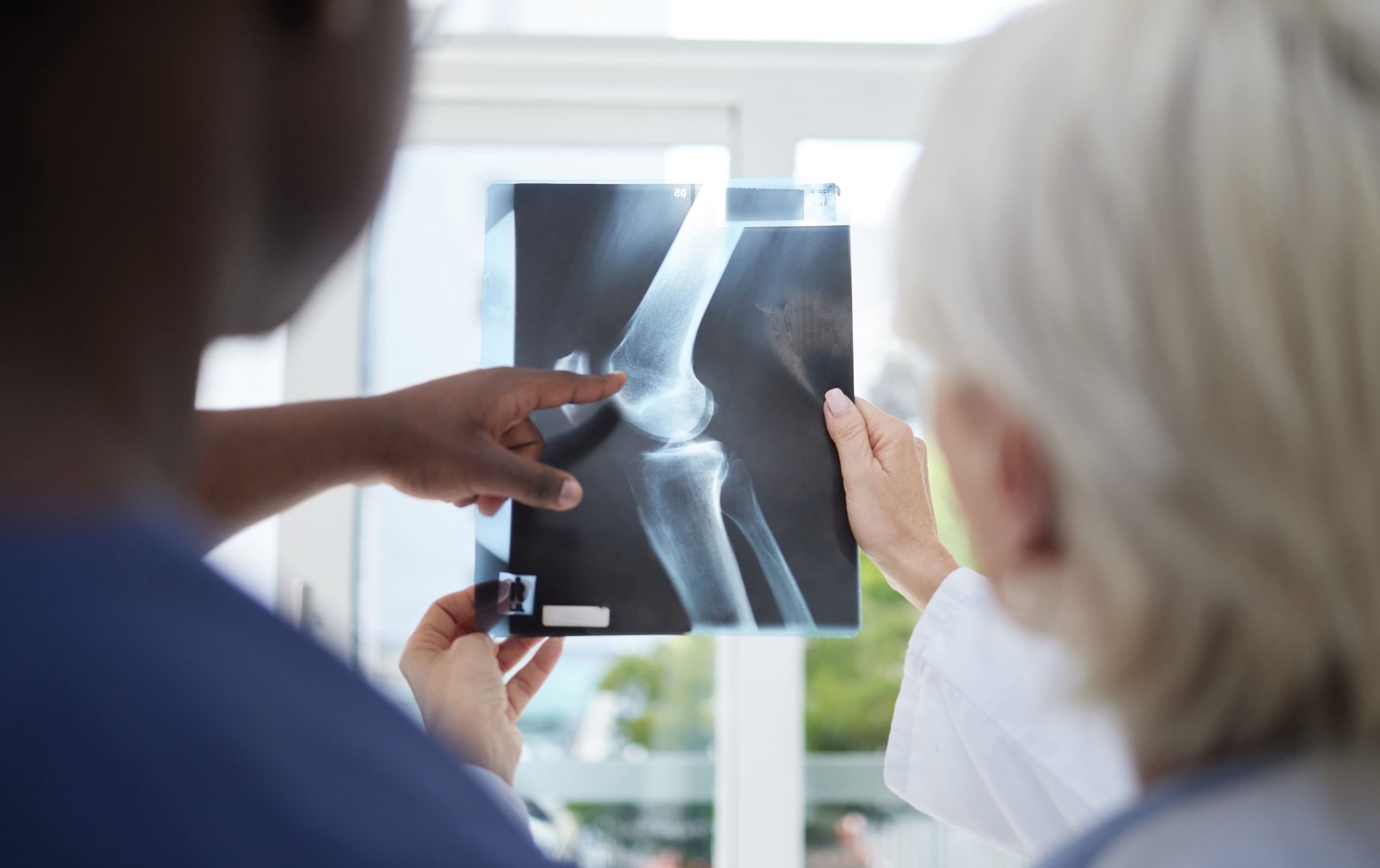
Imaging Studies
X-rays identify fractures, including avulsion injuries (where ligaments pull bone fragments from their attachments). Stress views—X-rays taken whilst applying force to the joint—reveal abnormal opening indicating ligament damage.
MRI (Magnetic Resonance Imaging, which uses magnets and radio waves to create detailed images of the inside of the knee) provides high-quality soft-tissue visualisation. It confirms ligament tears, grades their severity, and identifies associated meniscal or cartilage injuries. MRI findings guide treatment decisions, particularly regarding surgical versus non-surgical management and the optimal timing of any intervention.
Treatment Pathways
Non-Surgical Management
Many knee hyperextension injuries heal satisfactorily without surgery. Grade 1 (stretching without tearing) and grade 2 (partial thickness tears) injuries of most ligaments respond well to structured rehabilitation. The PCL, in particular, demonstrates reasonable healing potential even with complete tears, as the posterior capsule provides secondary restraint.
Rehabilitation progresses through phases:
- Protection and swelling control
- Range of motion restoration
- Strength rebuilding
- Functional training
Quadriceps strengthening receives particular emphasis, as this muscle group provides dynamic knee stability compensating for ligament laxity.
Bracing during activities may continue for several months. Functional braces with hyperextension stops prevent recurrent hyperextension during sports. They cannot fully replicate normal ligament function. Qualified healthcare professionals can provide personalised advice on treatment approaches tailored to your individual injury severity, activity level, and overall health profile.
Surgical Considerations
Complete ACL tears in active individuals typically require reconstruction (a surgical procedure in which the torn ligament is replaced with graft tissue) to restore stability for pivoting activities. Surgery involves replacing the torn ligament with graft tissue. This is commonly harvested from the patient’s hamstring tendons or patellar tendon, or obtained from donor tissue.
PCL reconstruction remains more controversial. Many surgeons recommend rehabilitation alone unless combined injuries create multidirectional instability. Complex injuries involving multiple ligaments require staged or combined reconstruction procedures.
Timing of surgery balances the benefits of early intervention against the risks of operating on swollen, inflamed tissues. Most surgeons prefer waiting until swelling subsides and range of motion returns—typically 2-4 weeks post-injury—before proceeding with reconstruction.
Rehabilitation and Recovery Timeline
Recovery timelines vary substantially depending on injury severity and the chosen treatment pathway. Minor hyperextension sprains resolve within 2-4 weeks with appropriate rehabilitation. Moderate injuries requiring extended bracing may take 6-12 weeks before returning to unrestricted activity.
ACL reconstruction involves a considerable rehabilitation period before returning to competitive sport. This extended timeline allows biological healing of the graft tissue within the bone tunnels and restoration of neuromuscular control (the coordination between nerves and muscles) patterns. Premature return risks graft failure and re-injury.
Rehabilitation milestones guide progression rather than arbitrary timelines. Achieving full range of motion, demonstrating adequate strength (comparable to the uninjured side), and passing functional testing permit safe progression of activity.
✅ Quick Tip
Maintain cardiovascular fitness during rehabilitation through low-impact activities. Swimming, cycling, or upper body exercises preserve conditioning without stressing the healing knee. This approach shortens the reconditioning phase once full activity resumes.
Long-Term Considerations
Some individuals develop chronic hyperextension laxity following initial injury, particularly if rehabilitation inadequately restores muscular control. This ongoing instability increases stress on joint surfaces, potentially accelerating cartilage wear over time.
Proprioceptive deficits (reduced awareness of joint position) persist in many cases despite successful structural healing. Continued balance and coordination training addresses these deficits. This reduces the risk of re-injury during challenging activities.
Activity modification may be necessary if persistent instability precludes safe participation in previous activities. Lower-demand activities, placing less rotational stress on the knee, often remain achievable even when high-level sport becomes impractical.
Preventing Knee Hyperextension
Neuromuscular training programmes (structured exercises that improve the coordination between your nerves and muscles) can help reduce knee injury rates in at-risk populations. These programmes emphasise proper landing mechanics. Bend the knees to absorb impact rather than landing with the knees locked. Regular participation during warm-ups provides cumulative protective benefits.
Hamstring strengthening helps control tibial position during deceleration, reducing hyperextension forces. The hamstrings act as dynamic ACL protectors. They pull the tibia backwards against the anterior translation forces.
Awareness of fatigue effects improves injury avoidance. Neuromuscular control deteriorates with exhaustion. This increases the likelihood of injuries late in games or training sessions. Appropriate conditioning and substitution patterns address this risk factor.
When to Seek Professional Help
- Inability to straighten or fully bend the knee after 48 hours
- Persistent instability or giving-way episodes during walking
- Swelling that increases rather than decreases over the first week
- Pain that prevents sleep or fails to improve with rest and ice
- Mechanical symptoms such as catching, locking, or clicking
- Numbness, tingling, or colour changes in the foot or lower leg
- Difficulty bearing weight after the first few days
Commonly Asked Questions
Can knee hyperextension heal on its own?
Minor hyperextension injuries involving stretching without structural tearing typically heal within several weeks with appropriate rest and rehabilitation. Complete ligament tears may stabilise through scar tissue formation. Functional instability often persists without surgical reconstruction in active individuals.
How do I know if I’ve torn my ACL from hyperextension?
A popping sensation at the time of injury, rapid swelling within two hours, and subsequent episodes of the knee giving way during pivoting activities suggest ACL damage. Clinical examination and MRI confirm the diagnosis. Some ACL tears produce surprisingly mild initial symptoms. A professional evaluation is warranted after any hyperextension injury.
Will I need surgery after hyperextending my knee?
Surgery depends on injury severity, affected structures, and activity goals. Isolated sprains and some complete PCL tears respond well to rehabilitation alone. Complete ACL tears typically require reconstruction for individuals wanting to return to pivoting sports. Your orthopaedic surgeon (a doctor who specialises in treating bones, joints, and related structures) can discuss specific recommendations based on imaging findings and functional requirements.
How long after knee hyperextension can I return to sports?
Return timelines range from 2-4 weeks for minor sprains to extended periods following ACL reconstruction. Achieving specific rehabilitation milestones—full motion, adequate strength, and successful functional testing—guides safe return rather than arbitrary time periods.
Can hyperextension cause permanent damage?
Severe hyperextension injuries may result in chronic instability, accelerated cartilage wear, or ongoing pain if inadequately treated. Appropriate acute management and thorough rehabilitation can help minimise long-term consequences. Even complete ligament tears achieve satisfactory outcomes in many cases with proper treatment.
Please note That Individual recovery experiences will vary due to personal health factors. This content is educational in nature and should not replace consultation with qualified healthcare professionals for tailored medical advice.

Experiencing Knee Pain or Injury?
Get a Personalised Treatment Plan
Find relief with our hip & knee specialists.
Make An EnquiryNext Steps
Knee hyperextension injuries range from minor sprains resolving within weeks to complex ligament damage requiring surgical reconstruction. Early assessment establishes accurate diagnosis, guides treatment selection, and optimises recovery potential. Rehabilitation milestones—full range of motion, adequate strength, and successful functional testing—determine safe return to activity rather than arbitrary timelines.
If you’re experiencing persistent knee instability, giving-way episodes, or difficulty returning to normal activities after a hyperextension injury, an orthopaedic surgeon can provide a comprehensive evaluation and discuss treatment options tailored to your activity goals.